All the key benefits you want
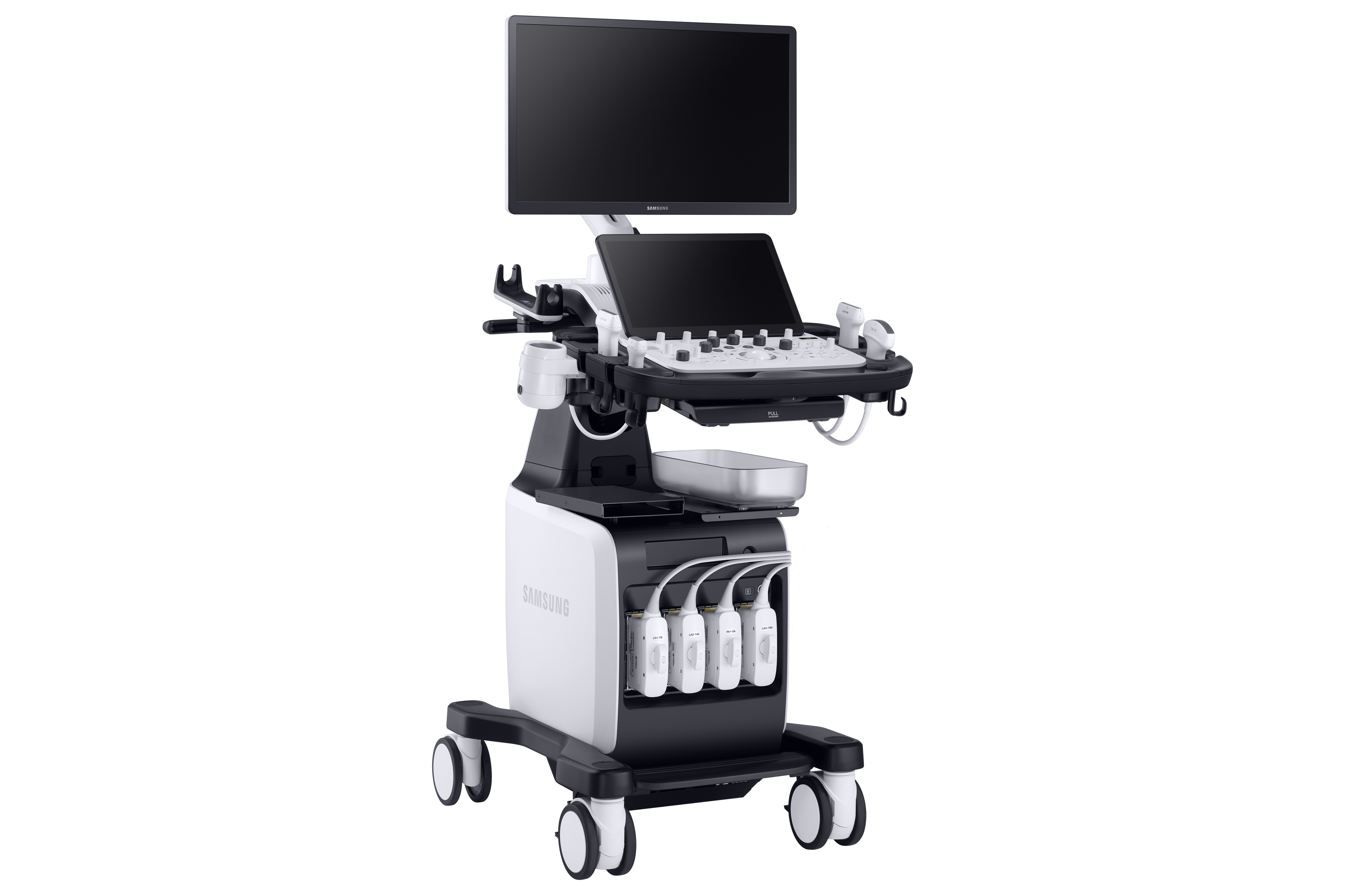
The V7 offers a fascinating performance and gives you the possibility to do what you want with comprehensive tools that feature the latest innovations.
For instance, EzHRI™, TAI™, and TSI™ are advanced abdominal dedicated diagnostic features, that help healthcare professionals make accurate clinical decisions by quantifying fatty liver in real time.
Rich in features, V7's versatile system is capable of a wide range of clinical applications that allow you to explore to the fullest.
Diagnose diverse and challenging clinical cases
The V7 comes with a variety of tools for diverse and challenging cases.
Healthcare professionals can execute targeted examinations with ease, using the necessary advanced features prepared in the right place.
Furthermore, various sophisticated 2D and color imaging features are supported for extraordinary image quality.
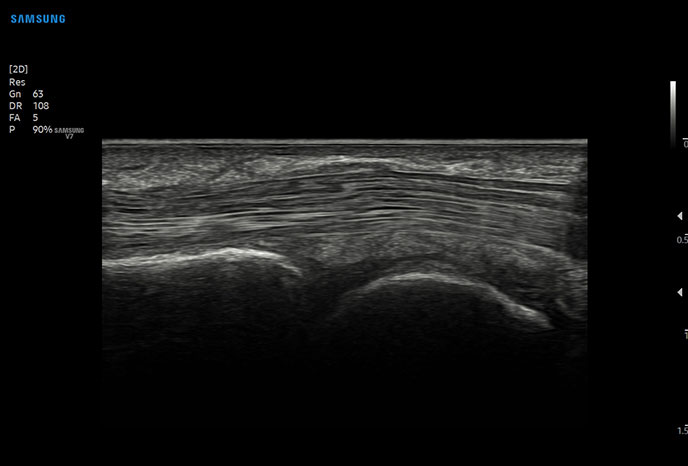
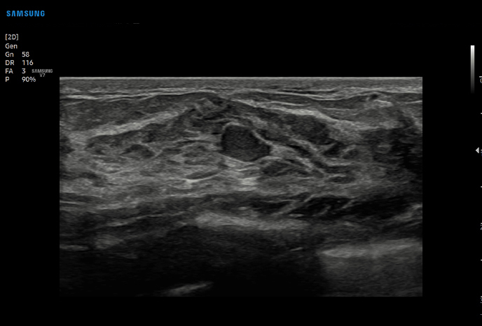
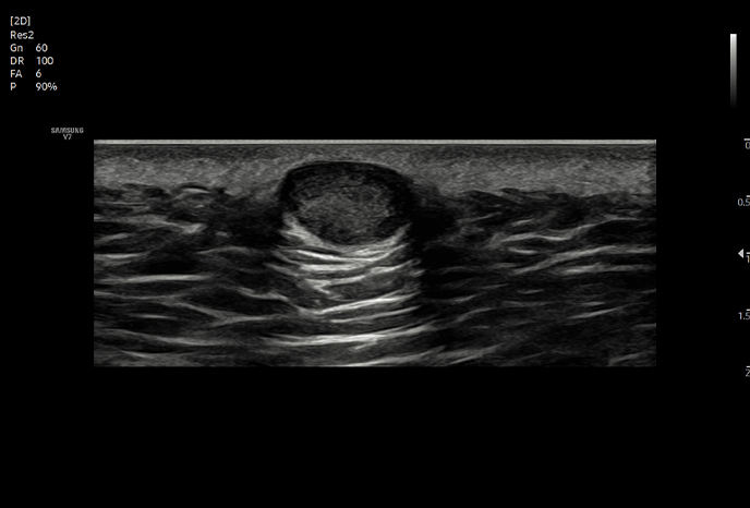
2D imaging
 |
 |
 |
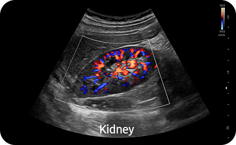
Color imaging
 |
 |
 |
Diagnostic features
 |
 |
 |
 |
 |
 |
 |
 |
 |
 |
![]()
Enriched diagnostic features with accuracy and precision
The V7 system comes with advanced features that assist in precise diagnosis and increasing throughput.
The V7’s variety of features and user-friendly interface aid in significantly improving the healthcare professionals’ daily ultrasound examination experience.
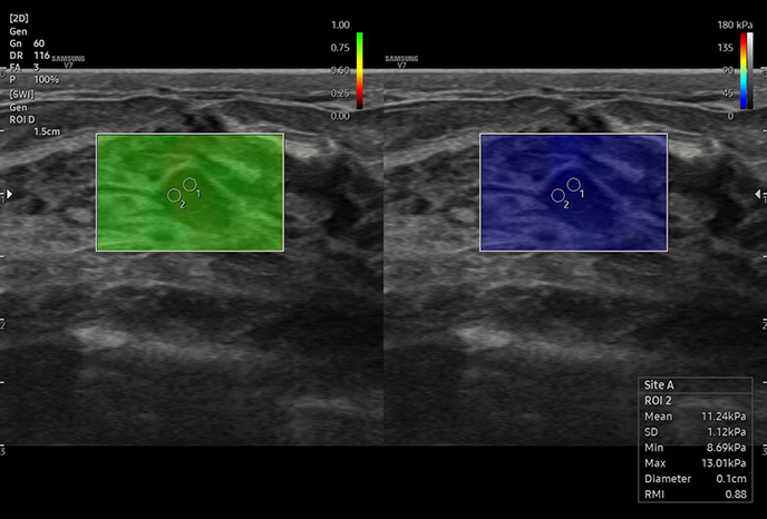
Display and quantify tissuestiffness in a non-invasive method
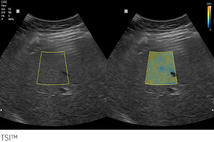
Quantitative measurement of liver fat with ultrasound signal
TSI™ (Tissue Scatter distribution Imaging) provides quantitative tissue scatter distribution measurement to assess steatotic liver changes.
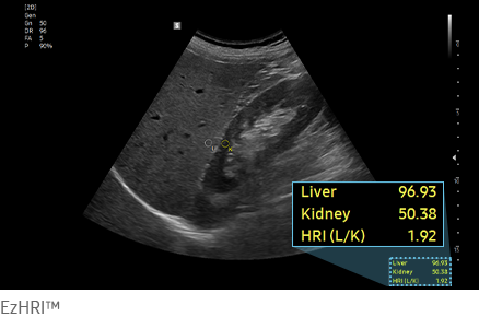
Hepato-renal index with automated ROI recommendation
EzHRI™ places 2 ROIs on the liver parenchyma and renal cortex and provides HRI ratio.
Perform multi-modality fusion biopsies with high precision
Contrast Enhanced Ultrasound
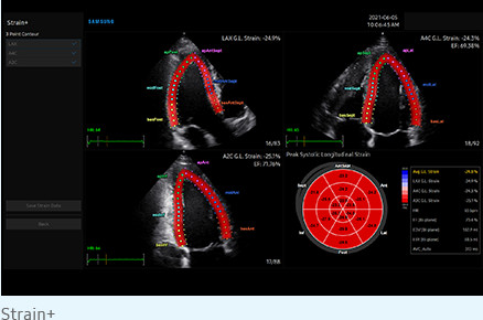
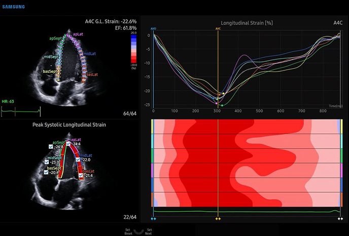
Quantify wall motion of the left ventricle
Score and report wall motion to determine heart and blood vessel function
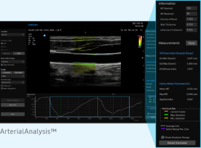
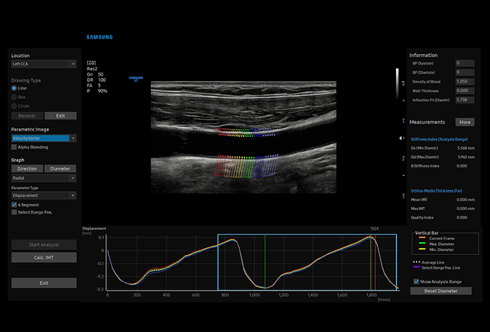
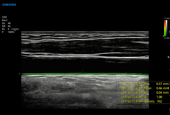
Detect functional changes of cardiovascular vessels
Measure IMT in one click
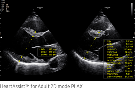
An automated reporting tool for heart diagnosis
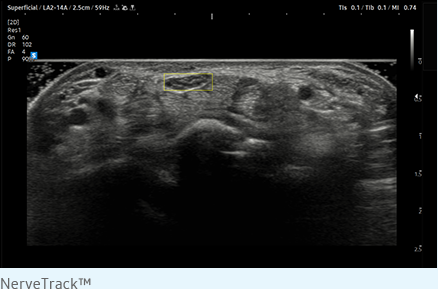
Detect and track nerves automatically with AI technology
Display needle tip clearly
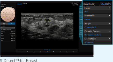
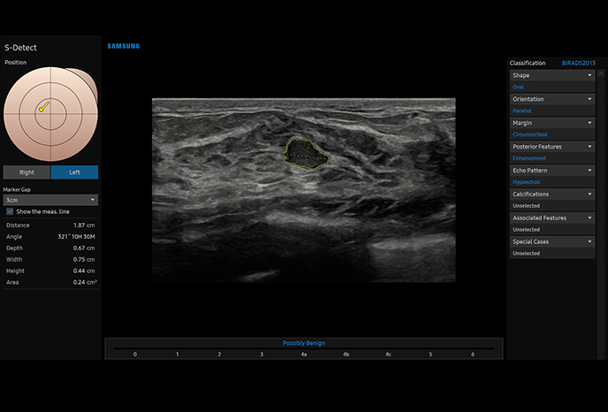
Analyze selected breast lesionsand report breast assessment
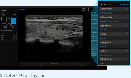
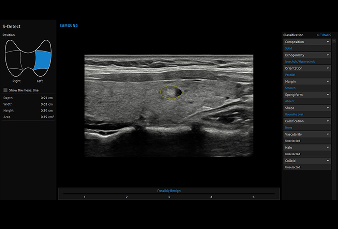
Analysis of selected thyroid lesion and reportingfor thyroid assessment
BTA: British Thyroid Association
EU-TIRADS: European Thyroid Imaging Reporting and Data System
K-TIRADS: Korean Thyroid Imaging Reporting and Data System
![]()
![]()
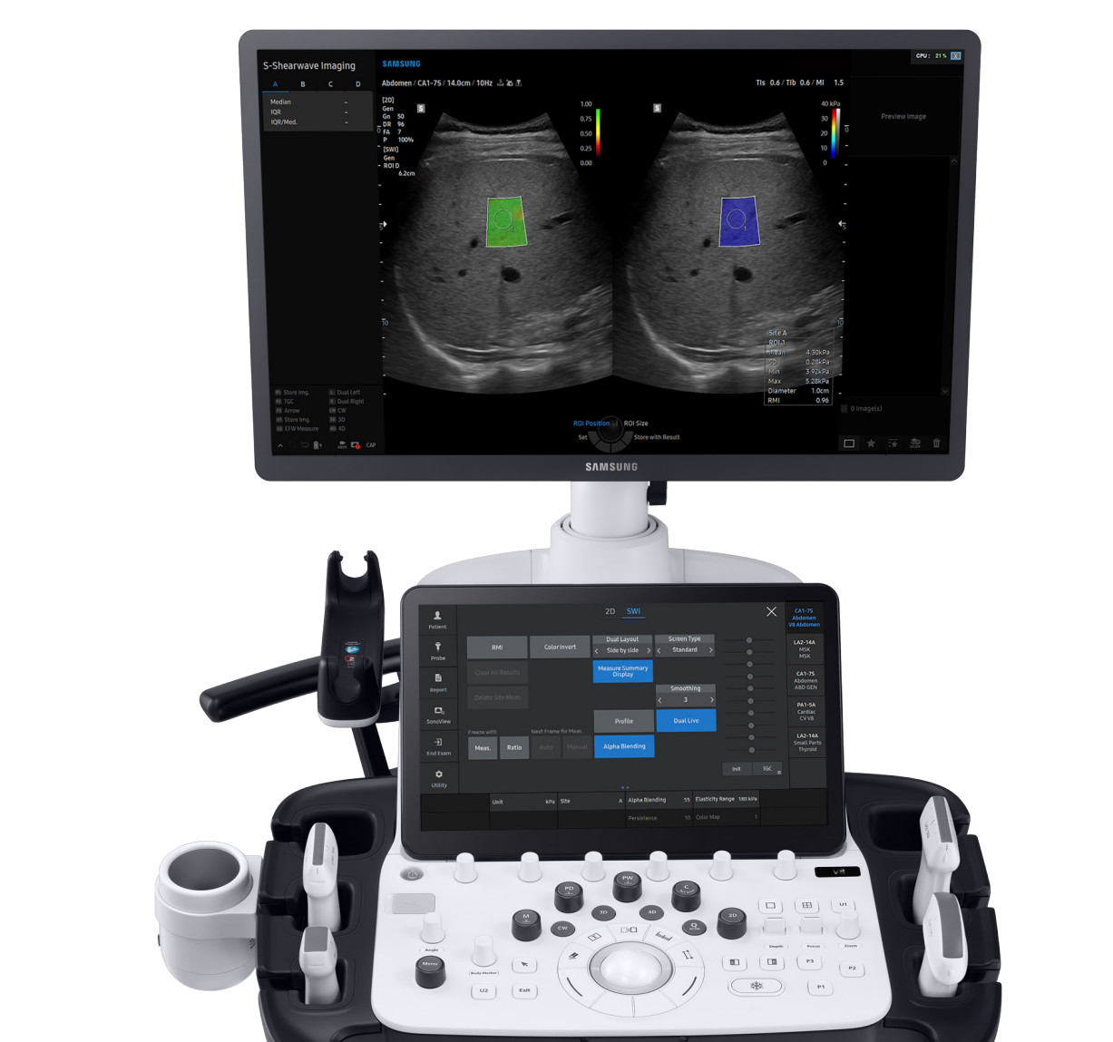
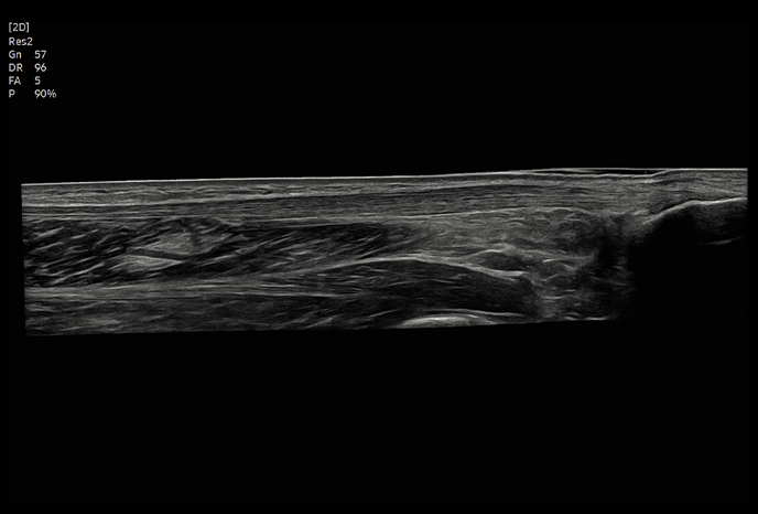
Extraordinary image quality delivers diagnostic confidence
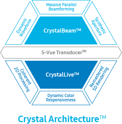 |
Gain insight into complex issues with exceptional image quality and resolution by Samsung’s core imaging engine, Crystal Architecture™. The proprietary technology combines enhanced 2D image processing and detailed color signal processing to optimize and refine the image. The cutting-edge V7 provides outstanding image clarity for a confident diagnosis. |
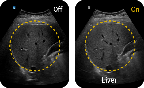
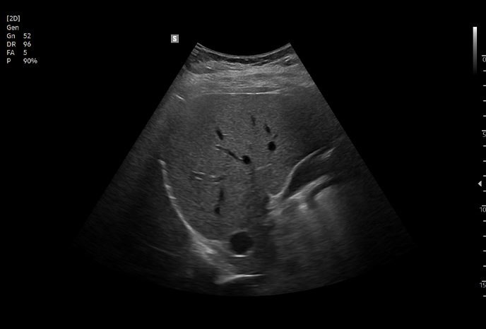
Enhance hidden structures in shadowed regions
ShadowHDR™ selectively applies high-frequency and low-frequency of ultrasound to identify shadow areas where attenuation occurs.
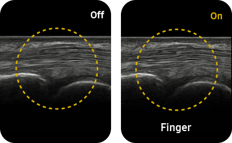
Clean up blurry areas in the image
HQ-Vision™ provides clearer images by mitigating the characteristics of ultrasound images that are slightly blurred than the actual vision.
Reduce noise to improve 2D image quality
ClearVision enhances the edge contrast and creates sharp 2D images for optimal diagnostic performance.
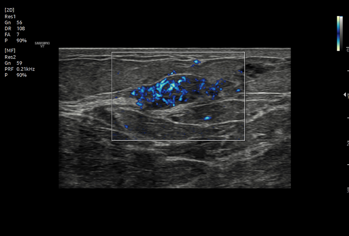
Visualize slow flow in microvascular structures
MV-Flow™ visualizes microcirculatory and slow blood flow to display the intensity of blood flow in color.
Show blood flow in vessels in a 3D like display
LumiFlow™ is a function that visualizes blood flow in 3 dimensional-like to help understand the structure of blood flow and small vessels intuitively.
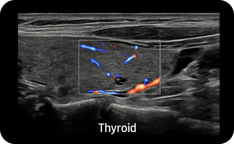
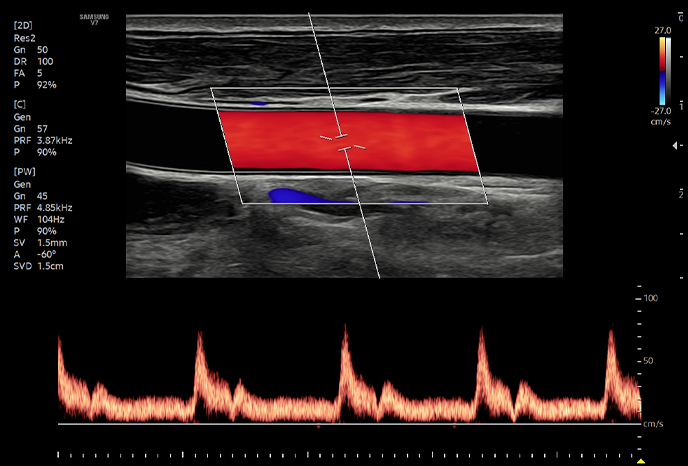
Examine peripheral vessels with directional power Doppler
S-Flow™, a directional Power Doppler imaging technology, can help to detect even the peripheral blood vessels. It enables accurate diagnosis when the blood flow examination is especially difficult.
![]()

 |
 |
 |
||
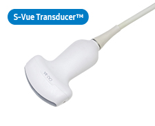
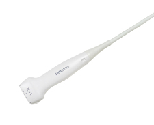
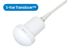
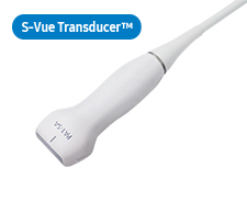
CA1-7SD*Application:Abdomen, Obstetrics, Gynecology, Pediatric, Musculoskeletal, Vascular, Urology, Thoracic |
CA3-10AApplication:Abdomen, Obstetrics, Gynecology, Pediatric, Musculoskeletal, Vascular, Urology, Thoracic |
CA4-10M*Application:Abdomen, Vacular, Pediatric |
 |
 |
 |
 |
|
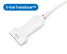
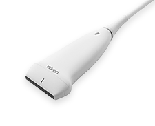
LA2-14AApplication:Small parts, Vascular, Musculoskeletal, Abdomen, Pediatric, Thoracic |
LA2-9AApplication:Small parts, Vascular, Musculoskeletal, Abdomen |
LA2-9S*Application:Small parts, Vascular, Musculoskeletal, Abdomen |
L3-22Application:Musculoskeletal, Small parts, Vascular, Pediatric |
|
 |
 |
|||
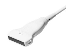
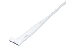
LA4-18AD*Application:Small parts, Vascular, Musculoskeletal, Abdomen |
LA3-22AIApplication:Small parts, Vascular, Musculoskeletal, Pediatric, Intraoperative |
 |
 |
|||
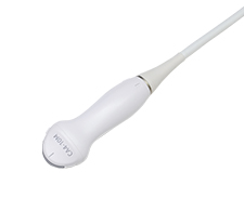
CV1-8ADApplication:Abdomen, Obstetrics, Gynecology, Urology |
EV2-10A*Application:Obstetrics, Gynecology, Urology |
 |
 |
 |
||
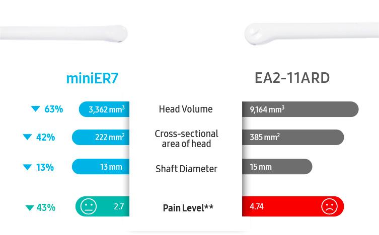
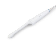
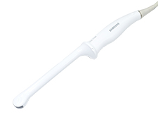
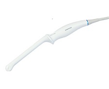
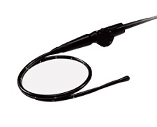
EA2-11ARD*Application:Obstetrics, Gynecology, Urology |
EA2-11AVD*Application:Obstetrics, Gynecology, Urology |
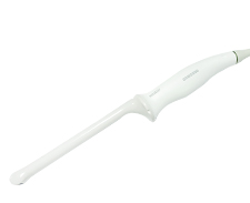
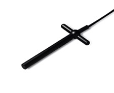
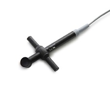
miniER7*Application:Obstetrics, Gynecology, Urology |
 |
 |
 |
||
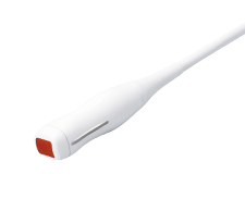
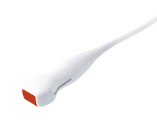
PA1-5APE*Application:Cardiac, Vascular, Abdomen, Pediatric, TCD, Thoracic |
PA4-12BApplication:Abdomen, Cardiac, Pediatric, Vascular, TCD |
PA3-8BApplication:Abdomen, Cardiac, Pediatric, Vascular, TCD |
 |
 |
|||
CW6.0Application:Cardiac |
DP2BApplication:Cardiac |
 |
||||
MMPT3-7Application:Cardiac |
* Ergonomic Transducer
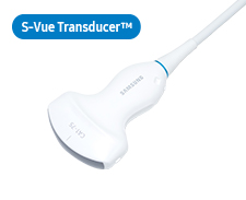
The new convex transducer design with a smooth and slim grip helps users to scan easily and comfortably.
The new endocavity transducer supports natural grip by moving the max width point to a more forward positionand also increased the length of the grip to allow balanced weight distribution.
Ultra Compact Prostate Ultrasound Transducer