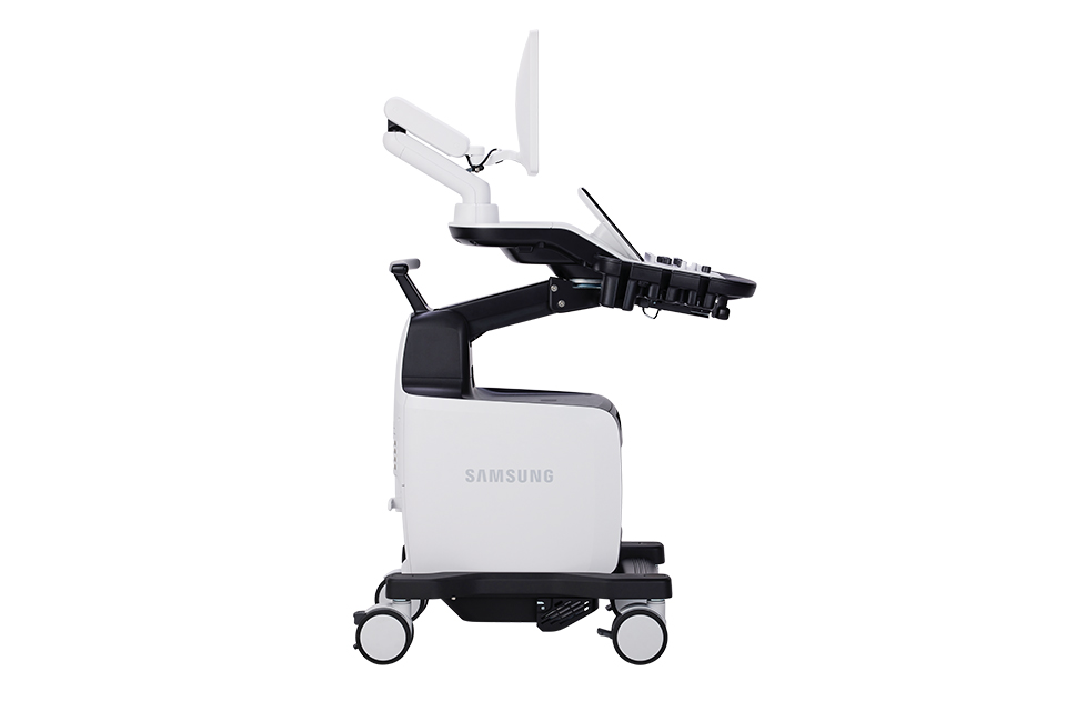
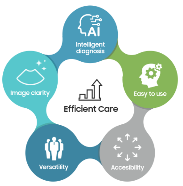
Embracing efficiency in your daily ultrasound scanning
Begin your journey towards efficient healthcare with the Samsung V6 ultrasound system. Our robust solution for general imaging offers both image clarity and advanced automated features. Additionally, Samsung’s cutting-edge imaging engine, Crystal Architecture™ ensures a reliable ultrasound experience.

 |
Experience simplicity with our easy-to-use system, specifically designed to alleviate your workload and enhance usability. Furthermore, our powerful system comes with battery capability, providing additional operational convenience. The Samsung V6 ultrasound system is a partner you can depend on to deliver exceptional efficiency to meet your daily ultrasound needs. |
![]()

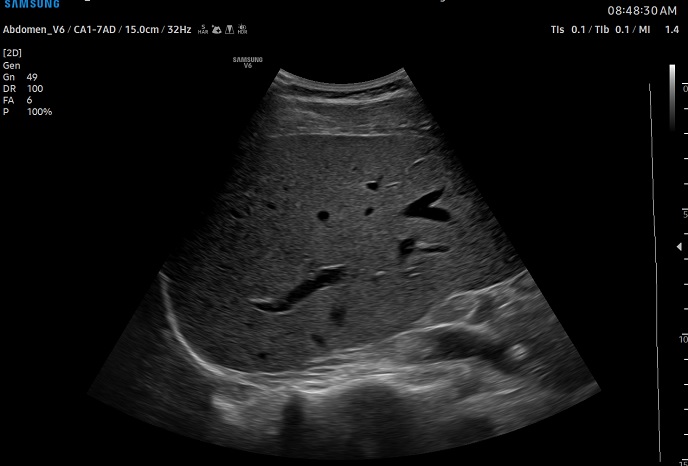
Elevating confidence with
superb imaging performance
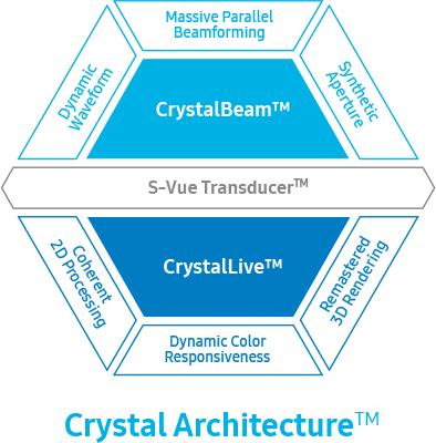 |
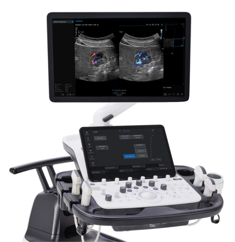
The V6 delivers exceptional 2D and color image quality tailored for general imaging, driven by Samsung’s core imaging engine, Crystal Architecture™. With its comprehensive imaging capabilities, the V6 is designed to seamlessly support your daily ultrasound scanning needs, enabling clear and accurate image acquisition. Experience confidence and accuracy in ultrasound scanning with the V6. |
2D imaging

Reduce noise to improve 2D image quality
ClearVision enhances the edge contrast and creates sharp 2D images for optimal diagnostic performance.
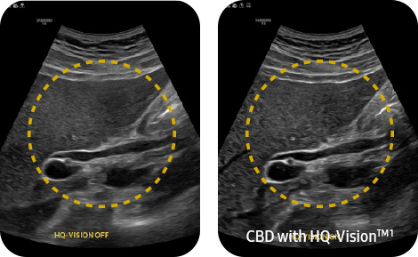
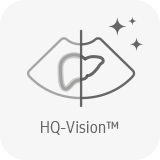
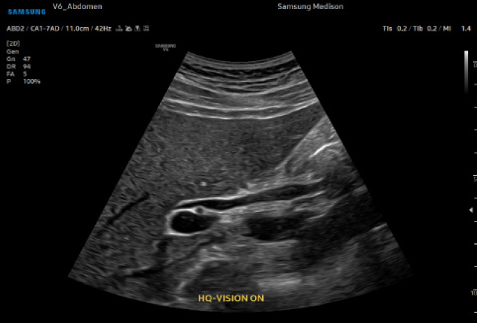
Clean up blurry areas in the image
HQ-Vision™ ¹ provides clearer images by mitigating the characteristics of ultrasound images that are slightly blurred than the actual vision.
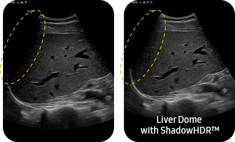
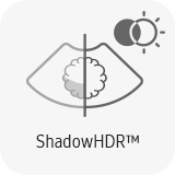
Enhance hidden structures in shadowed regions
ShadowHDR™ selectively applies high-frequency and low-frequency of ultrasound to identify shadow areas where attenuation occurs.
Color imaging
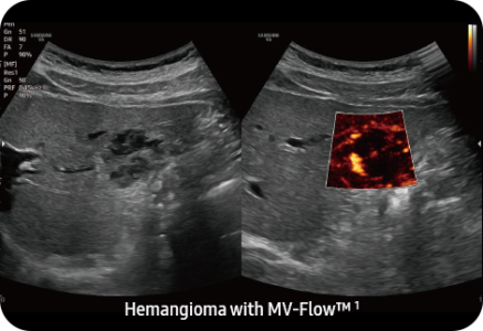
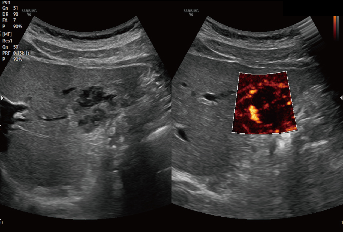
Visualize slow flow in microvascular vessels
MV-Flow™ visualizes microcirculatory and slow blood flow to display the intensity of blood flow in color.
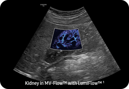
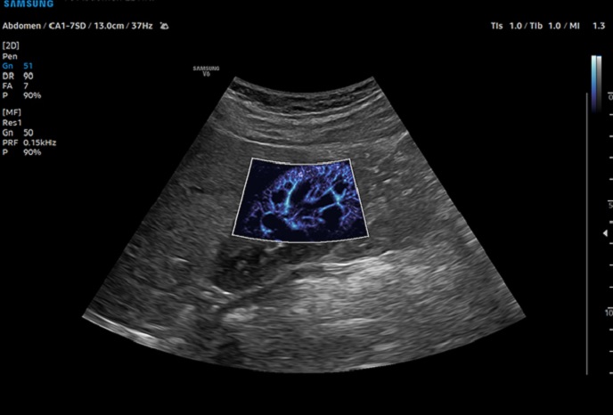
Show blood flow in vessels in a 3D-like display
LumiFlow™ is a function that visualizes blood flow in 3 dimensional-like to help understand the structure of blood flow and small vessels intuitively.
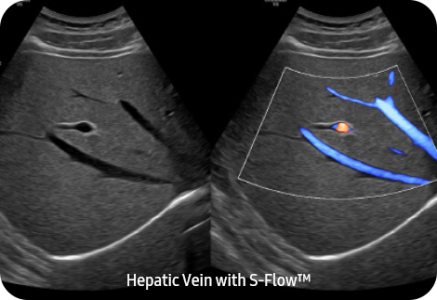
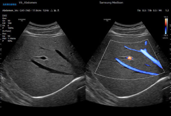
Examine peripheral vessels with directional power Doppler
S-Flow™, a directional Power Doppler imaging technology, can help to detect even the peripheral blood vessels. It enables accurate diagnosis when the blood flow examination is especially difficult.
Reach new diagnostic confidence
with comprehensive tools
Enhance your daily ultrasound diagnosis with the V6, a versatile solution created to efficiently support your clinical demands in general imaging. Benefit from our latest automation tools, which enable you to work with greater ease and achieve reliable results. Our aim is to assist you in prioritizing patient care,
and the V6 stands as an excellent choice.
![]()
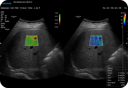
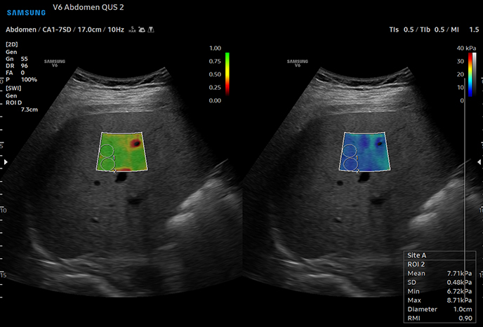
Display and quantify tissue stiffness in a non-invasive method
S-Shearwave Imaging™ allows the non-invasive assessment of stiff tissues in various applications. The color-coded elastogram, quantitative measurements, display options, and user-selectable ROI functions are useful for accurate diagnosis.
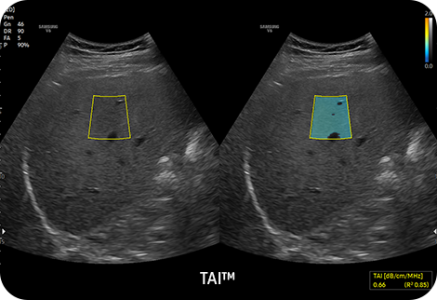
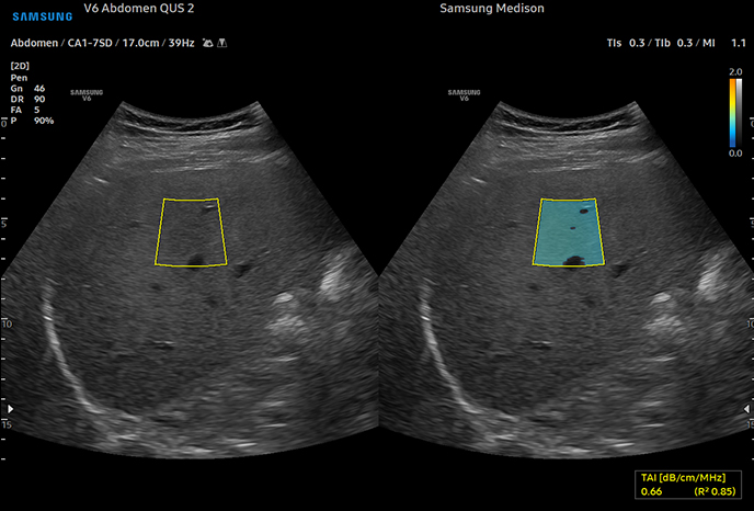
Quantitative measurement of liver fat with ultrasound signal
TAI™ (Tissue Attenuation Imaging) provides quantitative tissue attenuation measurement to assess steatotic liver changes.
TSI™ (Tissue Scatter distribution Imaging) provides quantitative tissue scatter distribution measurement to assess steatotic liver changes.
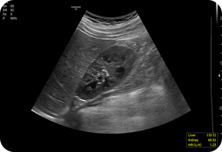
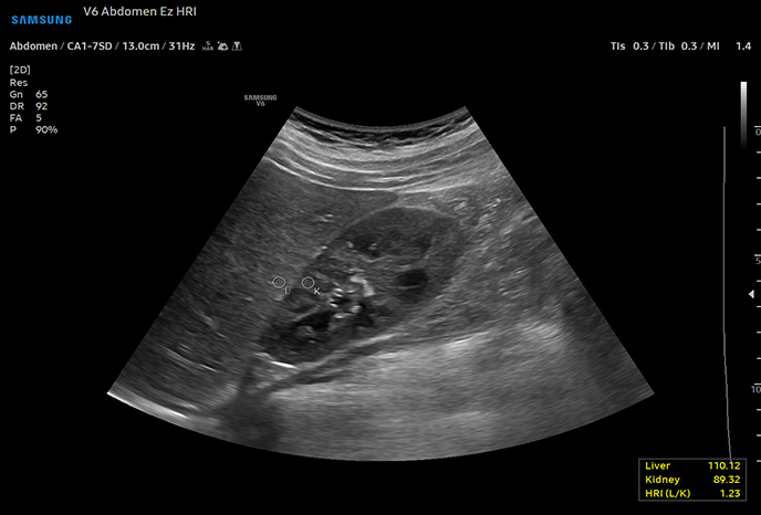
Hepato-renal index with automated ROI recommendation
HRI (Hepato Renal Index) is an index to quantify steatosis of a liver by comparing echogenicity between liver parenchyma and renal cortex. EzHRI™ places 2 ROIs on the liver parenchyma and renal cortex and provides HRI ratio.
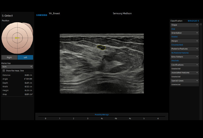
Analyze selected breast lesions and report breast assessment
S-Detect™ for Breast analyzes selected lesions in the breast ultrasound study and shows the analysis data, applies BI-RADS ATLAS* to provide standardized reporting; and helps diagnosis with the streamlined workflow
Analyze selected thyroid lesions and report thyroid assessment
S-Detect™ for Thyroid analyzes selected lesions in the thyroid ultrasound study and shows the analysis data, provides standardized reporting based on the ATA, BTA,EU-TIRADS, K-TIRADS, and ACR-TIRADS* guidelines; and helps diagnosis with the streamlined workflow.
- ATA: American Thyroid Association
- BTA: British Thyroid Association
- EU-TIRADS: European Thyroid Imaging Reporting and Data System
- K-TIRADS: Korean Thyroid Imaging Reporting and Data System
- ACR-TIRADS: American College of Radiology Thyroid Imaging Reporting and Data System
Display tissue stiffness in color image
A diagnostic ultrasound technique for imaging elasticity, ElastoScan+™ observes the transformation of the tissue strain by the internal or external forces, and converts relative stiffness into a color image.
Easy calculation of the strain ratio between two ROIs
E-Strain™ is designed to enable quick and easy calculation of the strain ratio between two regions of interest for day-to-day practice. Simply by setting the two targets, you can receive accurate, consistent results and make informed decisions in many types of diagnostic procedures.
Contrast Enhanced Ultrasound
CEUS+ is a contrast agent imaging technology. The micro-bubble contrast agent injected into the body through the vein or alike is subjected to perform nonlinear resonance due to stimulation of ultrasound energy.
An automated reporting tool for heart diagnosis
HeartAssist™, a feature based on Deep Learning technology, provides automatic classification of ultrasound image into measurement views required for heart diagnosis and provides measurement results.
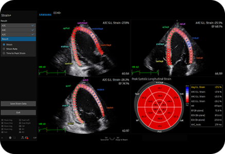
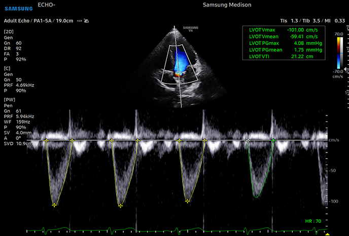
Quantify wall motion of the left ventricle
Strain+ is a quantitative tool for measuring global and segmental wall motion of the left ventricle (LV). Three standard LV views and a Bull’s Eye are displayed in a quad screen for easy assessment of the LV function.
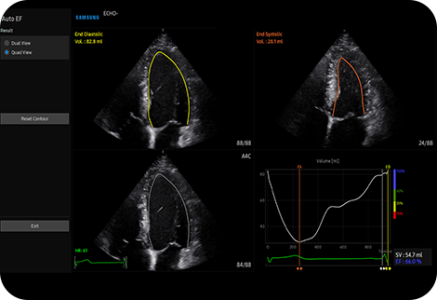
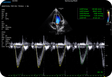
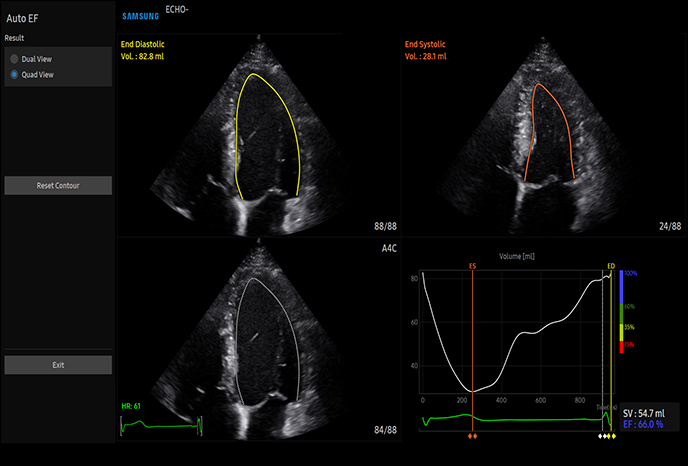
Measure ejection fraction of the left ventricle
AutoEF is a feature which conveniently measures and quantifies Ejection Fraction. By selecting the three points of the left ventricle, the volume at the end-systolic and end-diastolic points of the left ventricle is calculated, to assist in quick and efficient assessment of the heart function.

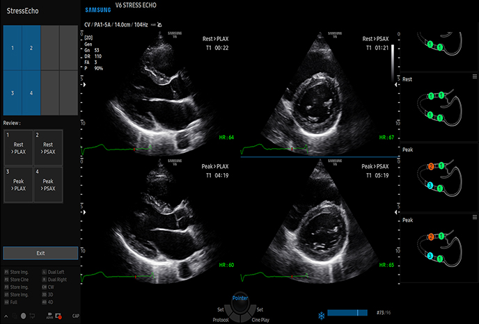
Score and report wall motion to determine heart and blood vessel function
StressEcho package includes wall motion scoring and reporting. It includes exercise StressEcho, pharmacologic StressEcho, diastolic StressEcho and free programmable StressEcho.
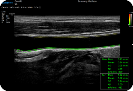
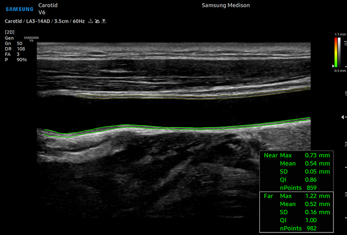
Measure IMT in one click
AutoIMT+ is a screening tool to analyze a patient's potential risk of cardiovascular disease. It allows easy intima-media thickness measurement of both the anterior and posterior wall of the common carotid by the click of a button.
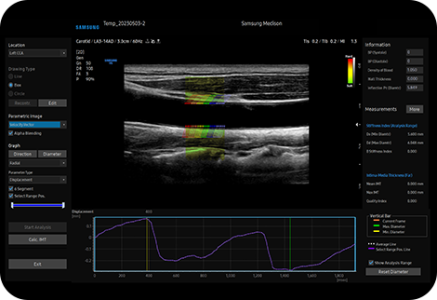
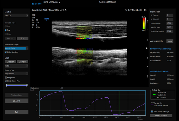
Detect functional changes of cardiovascular vessels
ArterialAnalysis™ detects functional changes of vessels, providing measurement values such as the stiffness, intima-media thickness and pulse wave velocity of the common carotid artery. Since the functional changes occur before morphological changes, this technology supports the early detection of cardiovascular disease.
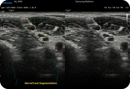
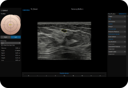
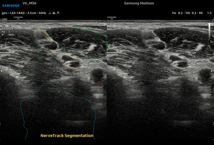
Detect and track nerves automatically with AI technology
NerveTrack™ , a feature based on Deep Learning technology, detects and provides information of the location of the nerve area in real-time during ultrasound scanning.
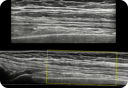
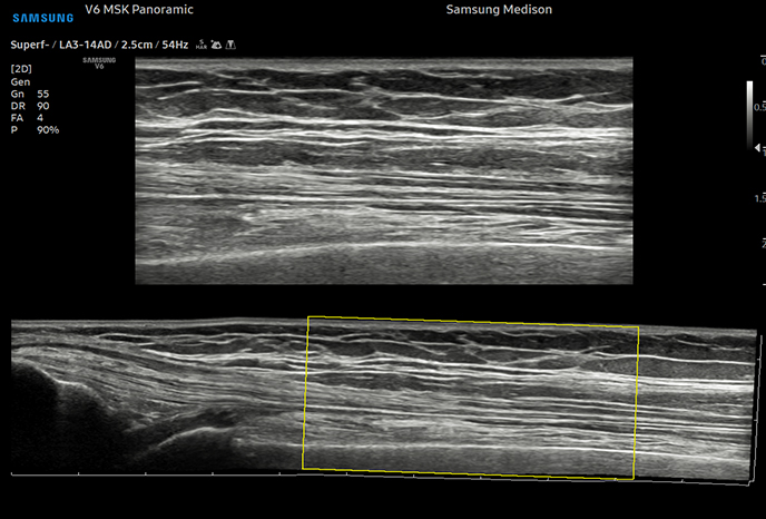
Display in extended field-of-view
Panoramic+ imaging displays as an extended field-of-view so users can examine wide areas that do not fit into one image as a single image. Panoramic+ imaging also supports angular scanning from linear transducer data acquisition.
Display needle tip clearly
With pinpoint precision, NeedleMate+™ delineates needle location when performing interventions such as nerve blocks. Improved accuracy and efficiency in the procedure are possible with beam steering added to NeedleMate+™.
![]()

 |
 |
 |
 |
|
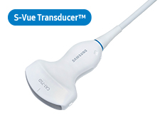
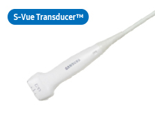
CA1-7ADApplication:Abdomen, Obstetrics, Gynecology, Musculoskeletal, Pediatric, Vascular, Urology |
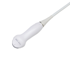
CA3-10AApplication:Abdomen, Obstetrics, Gynecology, Musculoskeletal, Pediatric, Vascular, Urology |
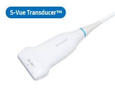
CA1-7SDApplication:Abdomen, Obstetrics, Gynecology |
CA4-10M*Application:Abdomen, Pediatric, Vascular |
 |
 |
 |
 |
|
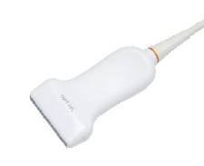
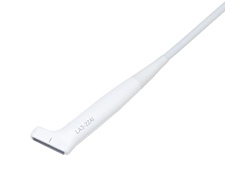
LA2-9S*Application:Small parts, Vascular, Pediatric, Musculoskeletal, Abdomen |
LA3-14ADApplication:Abdomen, Pediatric, Small parts, Vascular, Musculoskeletal |
L3-22Application:Musculoskeletal, Pediatric, Vascular, Small parts |
LA3-22AIApplication:Musculoskeletal, Intraoperative |
 |
 |
|||
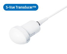
CV1-8AEApplication:Abdomen, Obstetrics, Gynecology |
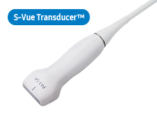
EV2-10A*Application:Obstetrics, Gynecology, Urology |
 |
 |
 |
||
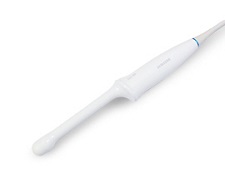
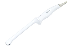
EA2-11ARE*Application:Obstetrics, Gynecology, Urology |
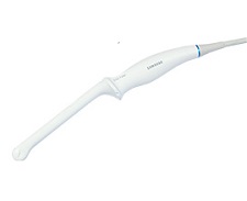
EA2-11AVE*Application:Obstetrics, Gynecology, Urology |
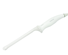
miniER7*Application:Obstetrics, Gynecology, Urology |
 |
 |
 |
||
PA1-5APEApplication:Cardiac, Vascular Abdomen, Pediatric, TCD, Thoracic |
PA4-12BApplication:Cardiac, Pediatric Abdoment, Vascular, TCD |
PA3-8BApplication:Cardiac, Pediatric, Abdomen, Vascular, TCD |
 |
 |
|||
CW6.0Application:Cardiac, Vascular, TCD |
DP2BApplication:Cardiac, Vascular, TCD |
 |
||||
MMPT3-7Application:Cardiac |
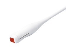
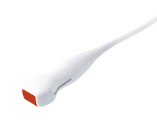
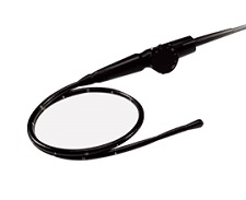
* Ergonomic Transducer (CA1-7S, EA2-11AR, EA2-11AV)
The new convex transducer design with a smooth and slim grip helps users to scan easily and comfortably.
The new endocavity transducer supports natural grip by moving the max width point to a more forward positionand also increased the length of the grip to allow balanced weight distribution.
![]()
![]()