- Description
- Specifications
Measure Known Tissue Elasticity
Shear wave elasticity imaging is an emerging biomarker with many possible applications, most prominently for determining the stage of liver fibrosis in a patient without the need for invasive biopsies. The design of the Shear Wave Liver Fibrosis Phantom, Model 039, was developed and validated in a joint study sponsored by the Quantitative Imaging Biomarker Alliance, and serves as the standard reference tool for determining sources of variance in shear wave elasticity measurements.
Tissue Equivalent Technology
The Model 039 consists of four phantoms – each filled with Zerdine® gel formulated with differing values of Young’s modulus, a tissue-average speed of sound of 1540 m/s and speckle contrast levels matching that of a healthy liver.
Designed to Comply with QIBA Standards
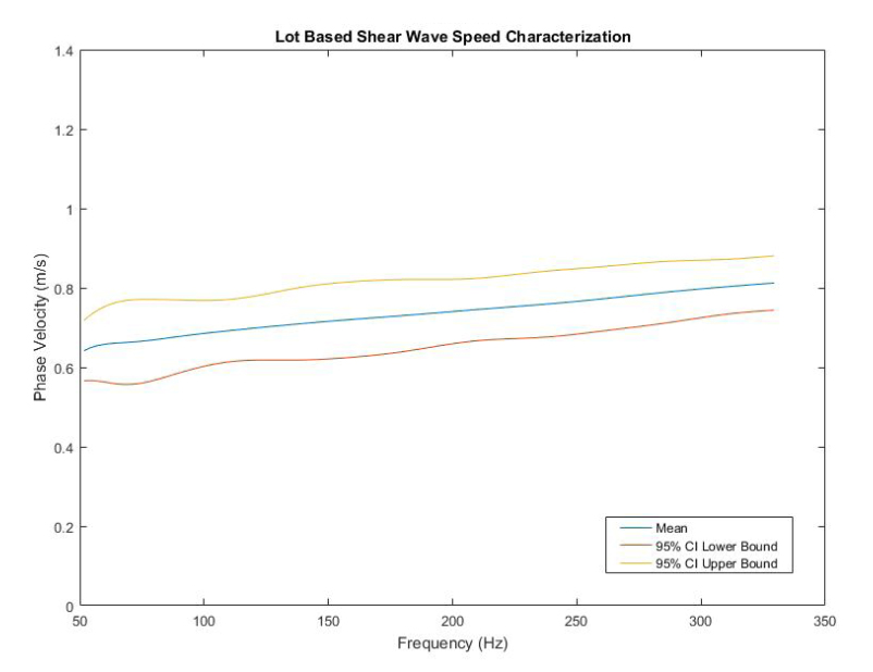
Certification of Young’s modulus will be provided with each phantom for proof of measurements with a precision of +/- 4%. Young’s modulus is tested on batch samples following ASTM standard D575-91 to ensure accurate elasticity. Density will also be measured to allow accurate conversion of shear wave speed to shear wave modulus and Young’s modulus.
Model 039 comes with a carry case for easy transport and phantom set up.
Key Features for Model 039
The model 039 set contains phantoms with Young’s Modulus Values spanning the range healthy livers to those with cirrhosis, as follows:
- Set of 4 phantoms, each with a different stiffness (Young’s modulus ranges from 2-36 kPa)
- Enables quantitative assessment of shear wave speed measurements used in the diagnosis of diffuse liver disease
- Certified measurement of shear wave speed according to protocol developed by Quantitative Imaging Biomarker Alliance Ultrasound Shear Wave committee
- Re-certification of phantoms available
Custom versions of this phantom, with different values for Young’s modulus and different sizes, are available upon special request.
|
|
||||||||||||||||||||||||||||||||||||||||||||||||||||||||||||||||||||||||||||||||||

