- Description
- Specifications
Head Phantom (mixed brain lesions)
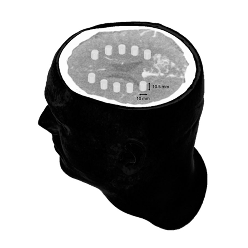
This head phantom can be used in CT for evaluation of low-contrast signals in the brain. It was designed to enable evaluation of diagnostic software, including AI tools.
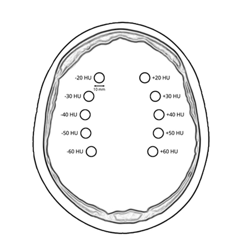
The phantom simulates a contrast medium enhanced head in arterial phase (CT angiography) and has 10 low-contrast lesions in the centrum semiovale. The neck is included up to the fifth cervical vertebra.
The phantom provides a detailed and realistic simulation of soft and bone tissue, including small details such as lymph nodes. The right hemisphere has an arteriovenous malformation. Air voids are filled with a cellulose-polymer composite of approx. -80 HU.
The phantom can be used for detection, segmentation and classification tasks and other common methods of image quality evaluation.
|
|

Head Phantom (mixed brain lesions)
This head phantom can be used in CT for evaluation of low-contrast signals in the brain. It was designed to enable evaluation of diagnostic software, including AI tools.
The phantom simulates a contrast medium enhanced head in arterial phase (CT angiography) and has 10 low-contrast lesions in the centrum semiovale. The neck is included up to the fifth cervical vertebra.
The phantom provides a detailed and realistic simulation of soft and bone tissue, including small details such as lymph nodes. The right hemisphere has an arteriovenous malformation. Air voids are filled with a cellulose-polymer composite of approx. -80 HU.
The phantom can be used for detection, segmentation and classification tasks and other common methods of image quality evaluation.
|
|


The Alderson Radiation Therapy Phantom
Accurate Organ Dosimetry Measurements with Minimal Dosimeters
- Approximately 10,000 phantoms in use all over the world for 30 years
- Indispensable quality-assurance tool
- Molded of tissue-equivalent material
The Alderson Radiation Therapy (ART) Phantom is a refined and improved version of the Alderson RANDO Phantom in both design and materials. ART Phantoms are designed within highly sophisticated technological constraints and follow ICRU-44 standards. Additionally, they provide integrated tests of the entire chain of treatment planning and delivery.
ART Phantoms are molded of tissue-equivalent material and designed for accuracy and ease of use.
Anatomy
The male ART represents a 175 cm (5 ft. 9 in.) tall, 73.5 kg (162 lb.) male, and the female ART represents a 155 cm (5 ft. 1 in.) tall, 50 kg (110 lb.) female.
The ART Phantom is transected-horizontally into 2.5 cm thick slices. Each slice has holes which are plugged with bone-equivalent, soft-tissue-equivalent or lung tissue equivalent pins which can be replaced by TLD holder pins. Holder pins may be ordered separately.
Soft-tissue-equivalent coatings produce slices with glass smooth interfaces. These coatings are cut away over the air spaces of the oronasal pharynges, trachea, and stem bronchi. Dosimetry holes are drilled in grids 3 cm x 3 cm or 1.5 cm x 1.5 cm in 5 and 7 mm diameters thereby allowing for detailed measurements of dose distributions.
Breast Attachments
Breasts are available in various sizes. They can be sliced in frontal planes, drilled or undrilled for film dosimetry. Slices can receive any of the pins listed in the TLD Dosimeters and Fittings section, below. Breasts of male and female ART Phantoms are contoured to blend realistically with the thoraxes and attached with nylon screws. The male chest with attached breasts serves as a large female.
Materials
Soft Tissues: There are unlimited, small variations in density and absorption throughout the human body. Phantom soft tissue is closely controlled to have the average density of these tissues.
Skeletons: RSD skeletons are highly detailed polymer moldings which reproduce the shape, mass density and attenuation coefficients of cortical bone and spongiosa. This allows for continuous production of phantoms, instead of the sporadic production required by the limited availability, variable size and uncertain chemical composition of human skeletons. Our proprietary moldings allow for continuous production, eliminate the restrictions of human skeleton bones (including limited availability, variable size, and uncertain chemical composition), and avoid the loss of marrows in dried natural skeletons thereby making RSD skeletons superior to “real bone.”
Molds for the RSD cortical bone and spongiosa were made from human skeletons consistent with the sizes of the soft tissue molds.
RSD skeletons conform closely to the standards established by the International Commission on Radiation Units and Measurements (ICRU Report No. 44); mass density is reduced slightly to take into account a small decrease in calcium content for older patients.
Lungs: Lungs are molded from syntactic foam, with a specific gravity of 0.30 g/cc.
TLD Dosimeters and Fittings
Phantoms are shipped with all dosimetry holes filled with blank pins. Pins for TLD chips have recesses at one end measuring 3.2 x 3.2 x 0.9 mm. Pins for TLD rods have 1 mm-diameter holes cross-drilled at the centers of the pins. All pins are 2.50 cm long unless otherwise specified. Pins may also be ordered to accommodate various types of OSLD dosimeters. Tissue equivalent plugs specifically machined for TLD chips, TLD rods, TLD bars, TLD cubes, MOSFET detectors, as well as LANDAUER® OSL MicroSTAR® and nanoDot® holders, are also available.
LANDAUER®, MicroSTAR®, and nanoDot® are registered trademarks of LANDAUER®, a division of Fluke® Corporation.
Assembly
ART Phantom slices are held between aluminum plates by nylon tie rods. Knobs at the end of the rods clamp the slices tightly in proper alignment. Both internal and external assembly devices are included.
The external assembly facilitates film dosimetry, while the internal assembly is used generally with TLDs or ion chamber dosimetry.
|
Order Requirements
Product Gender:
Volume:
Sliced or Unsliced:
Hole Grid:
Hole Size:
Side:
Example:
Model Numbers
|

This child phantom has different lifelike densities for tissue, bones and lungs. It is cut into slices and has holes for dosimeters.
The size is 60cm. Delivery with holding and fixation frame.
|
|

This phantom is developed for the treatment planning and machine adjustment in the radiation therapy. The body consists of 3 cm slices with a 3 x 3 cm hole matrix for inserting glass/TLD dosimeters.
The model material has a natural radio transparency allowing the correct adjustment of the machines. This makes it ideal for planning and machine adjustment. The phantom has a holding and fixation frame which allows to position the phantom exactly.
|
|