- Description
- Specifications
|
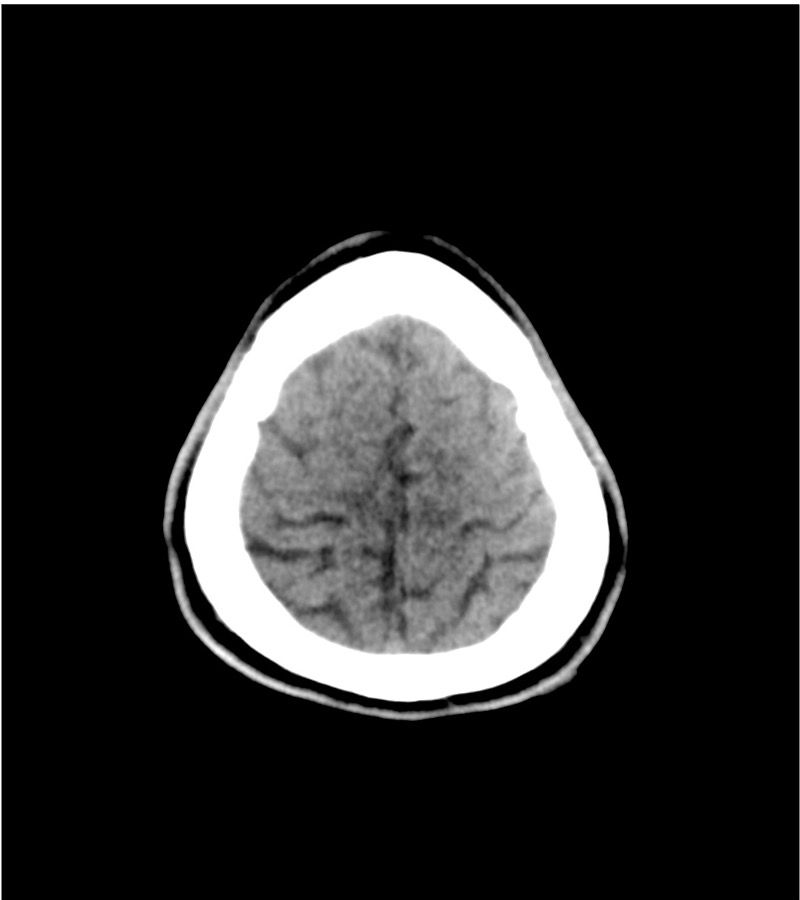
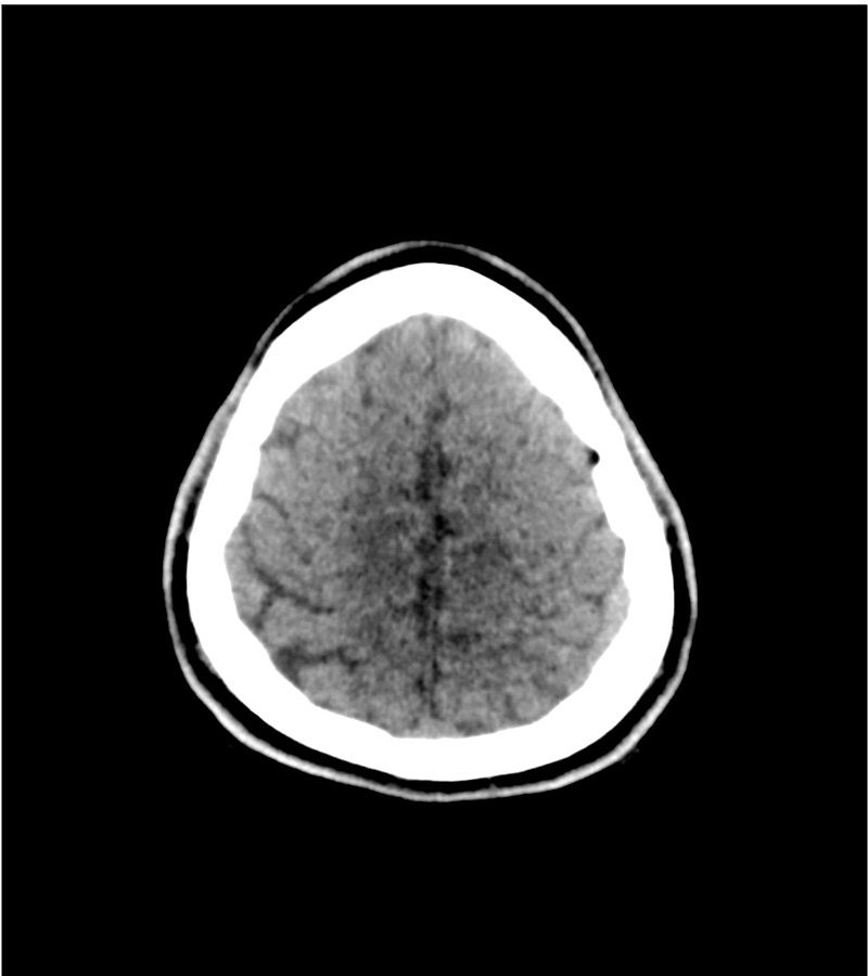
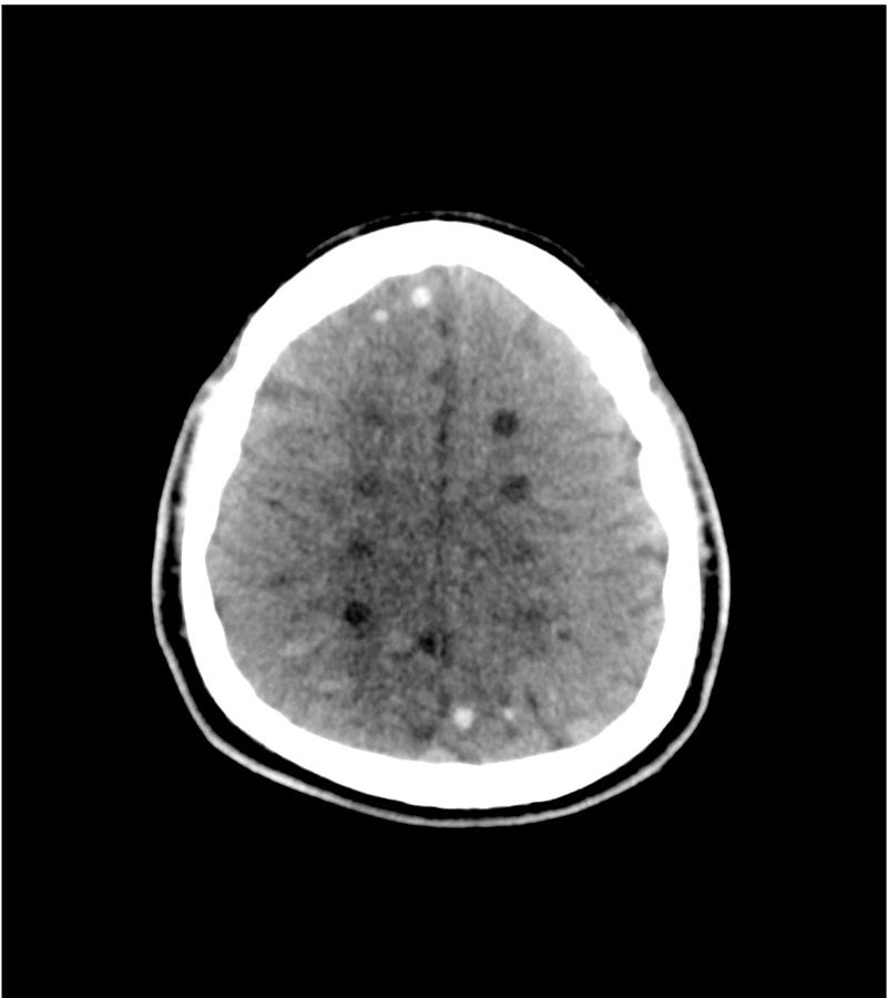
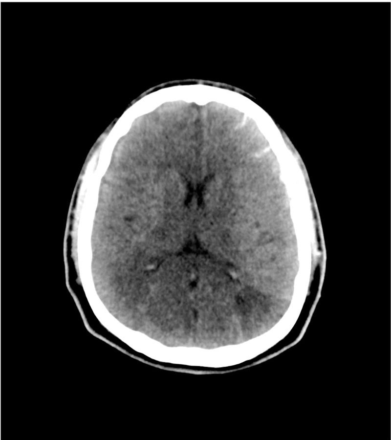
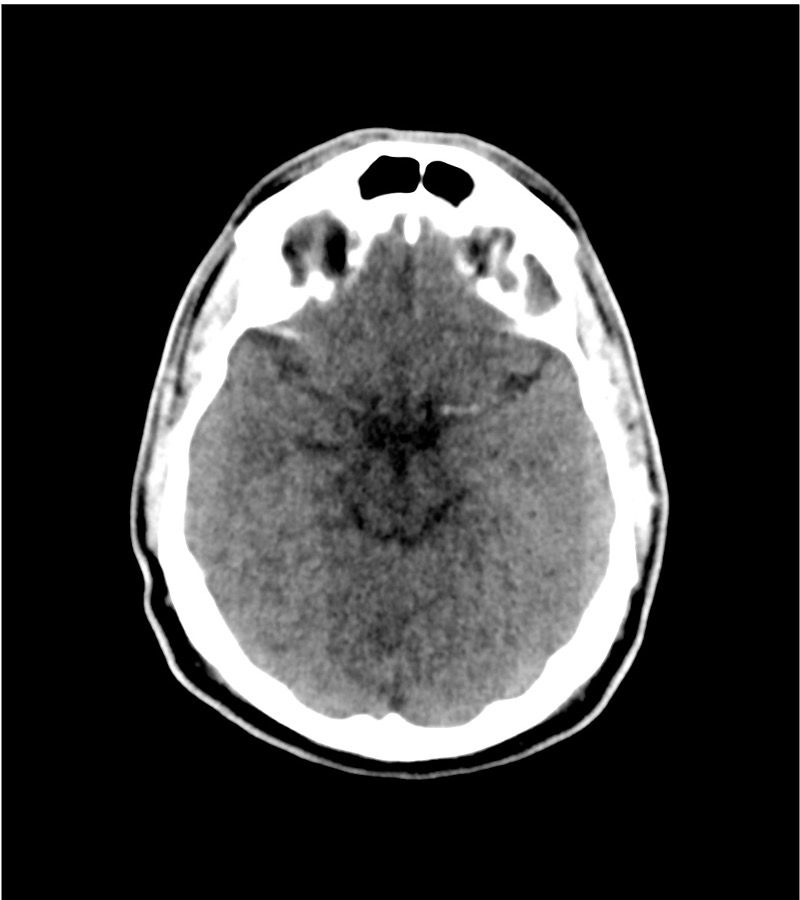
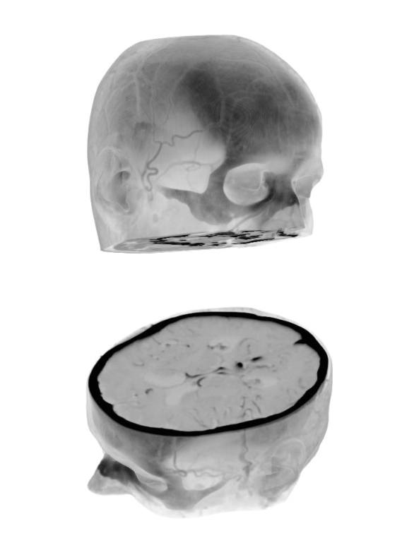
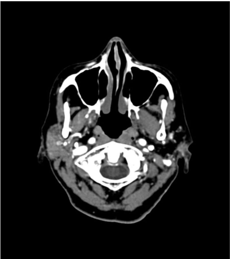
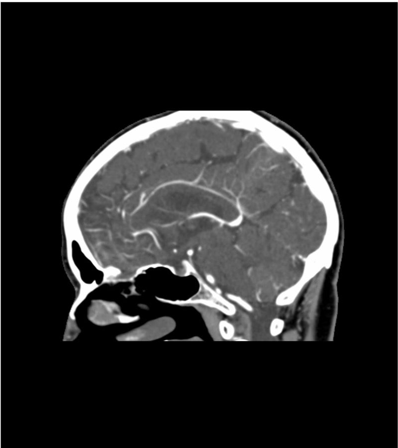
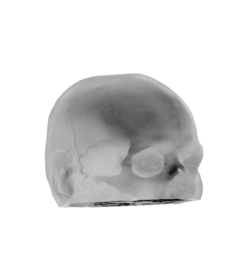
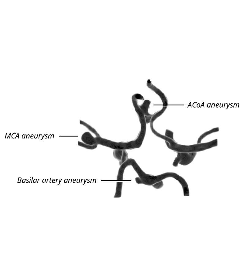
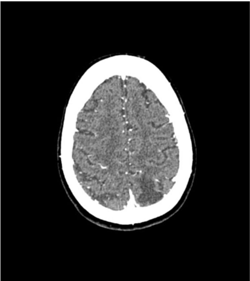
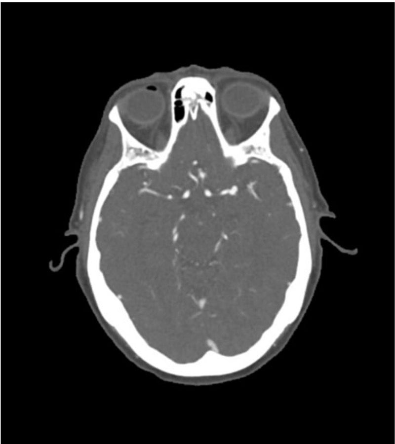
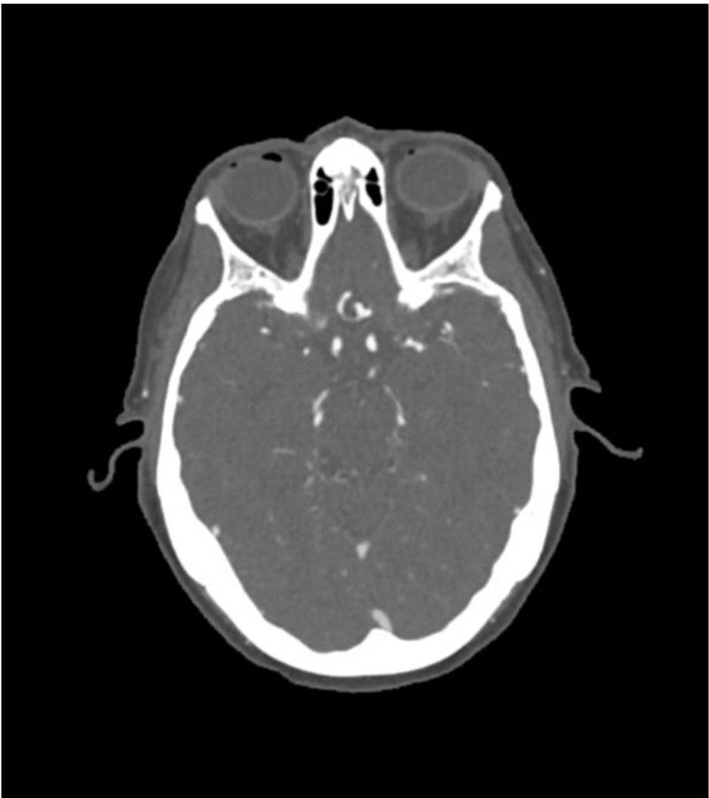
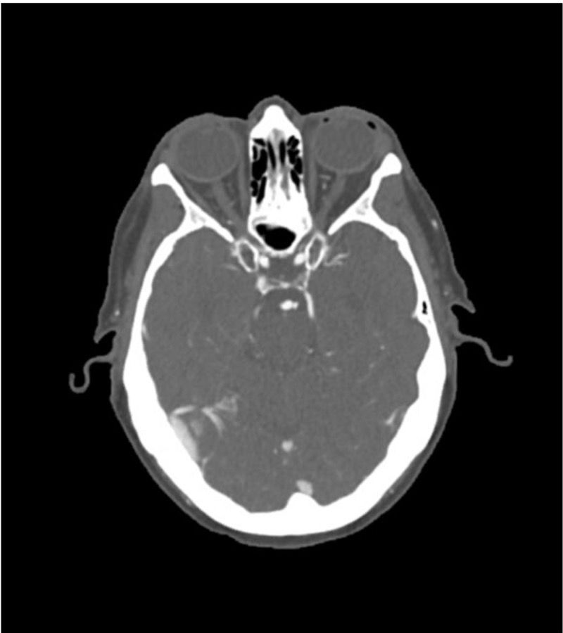
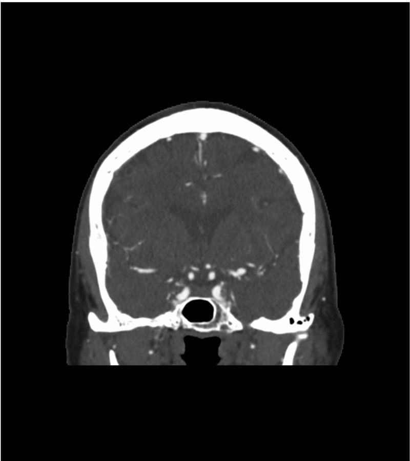
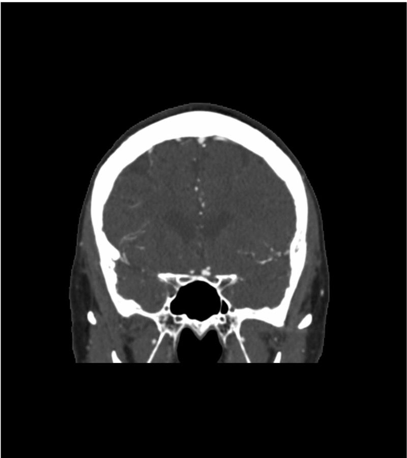
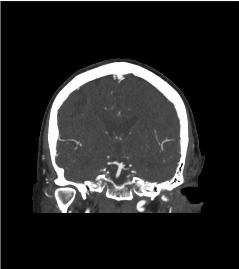
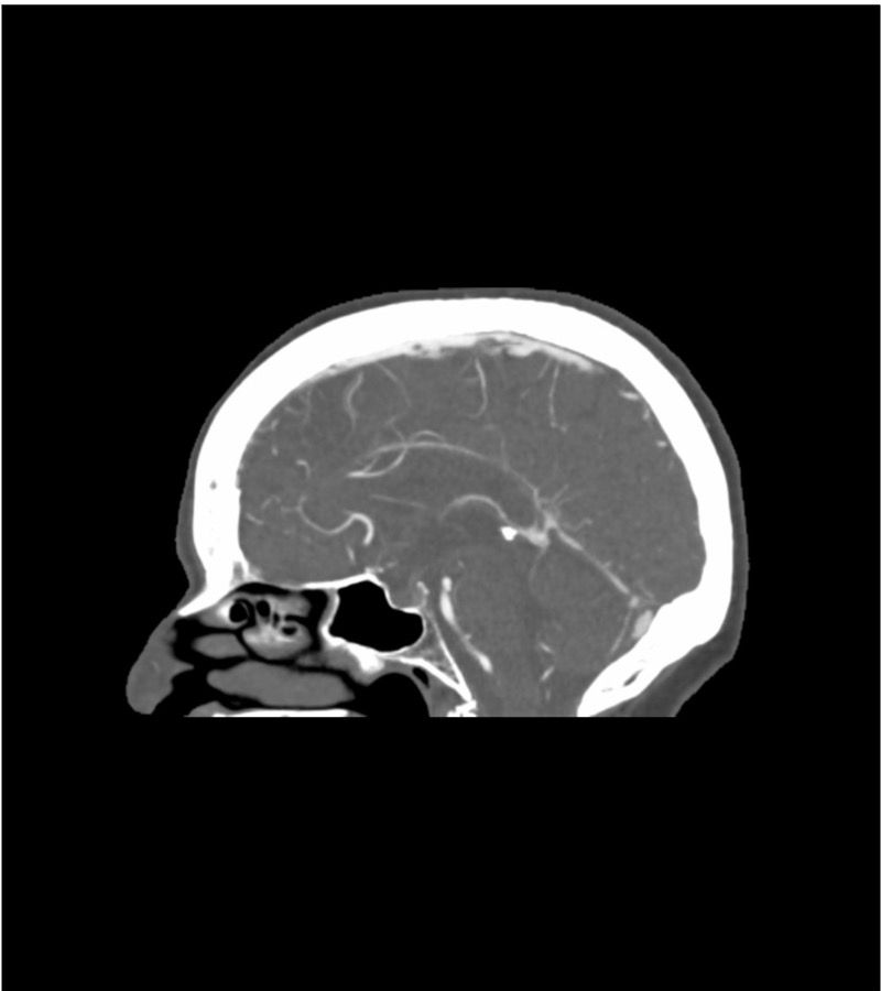
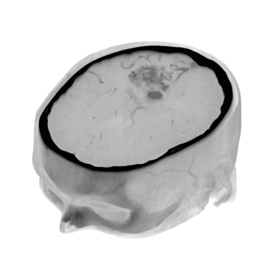
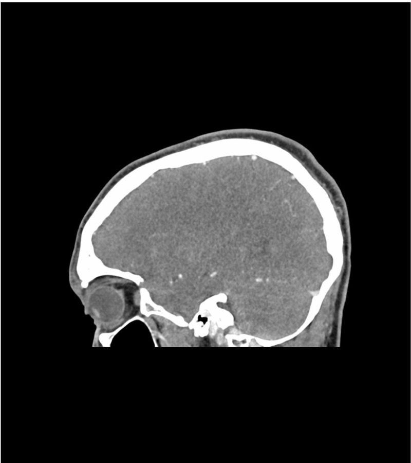
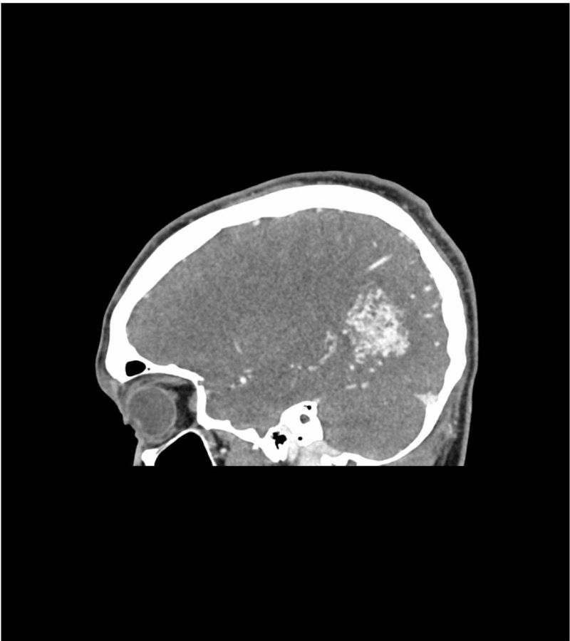
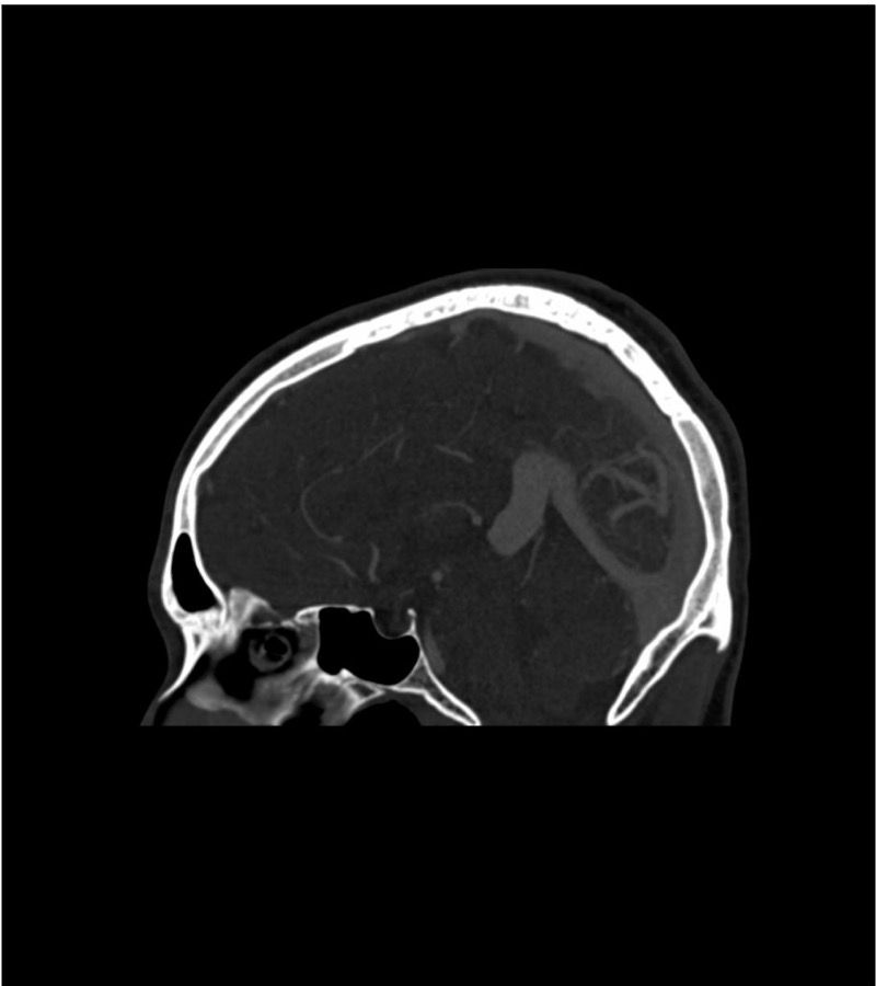
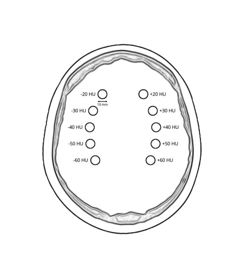
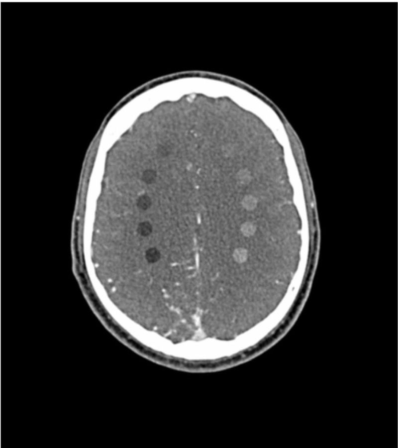
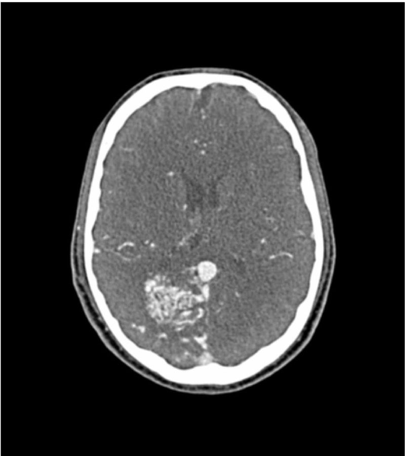
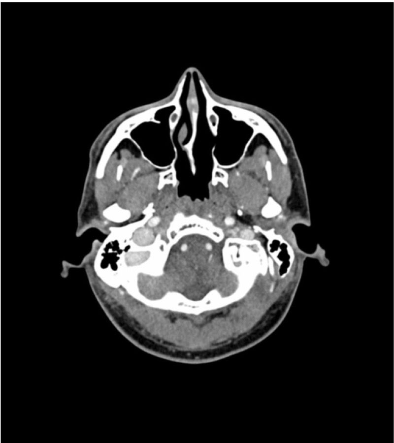
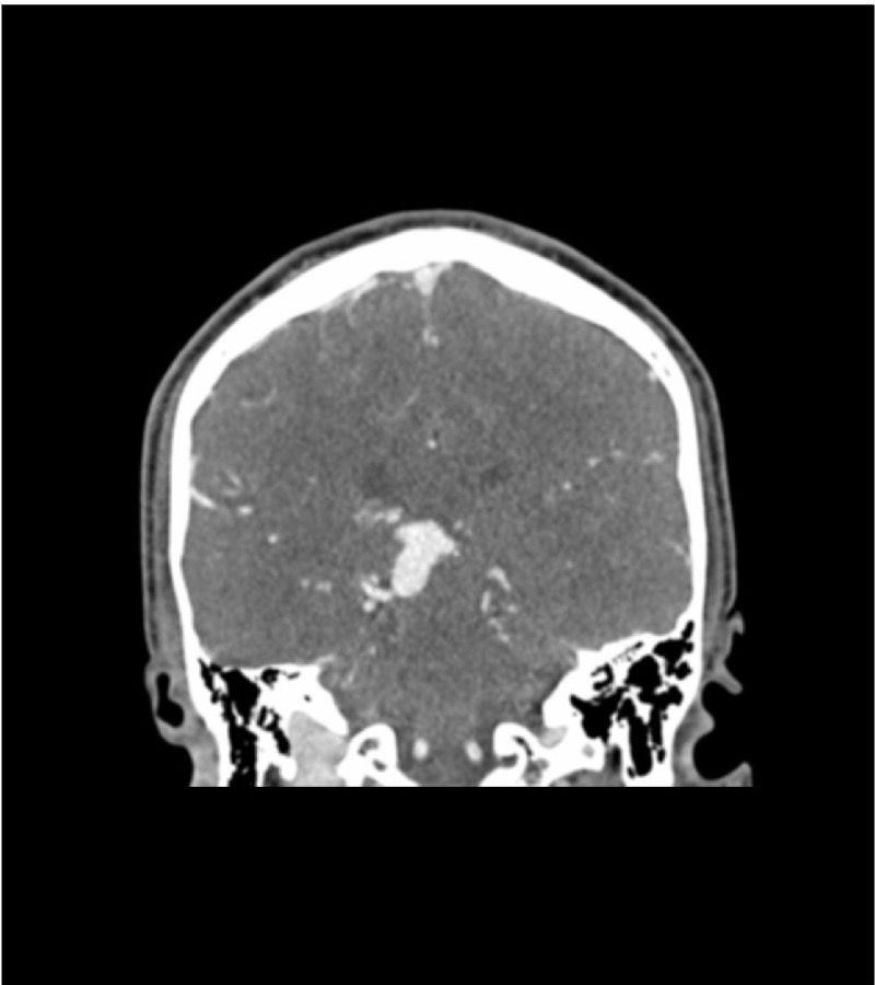
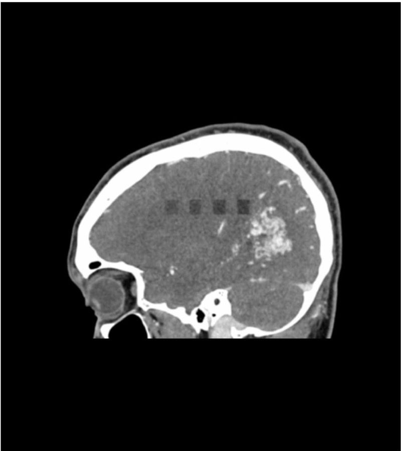
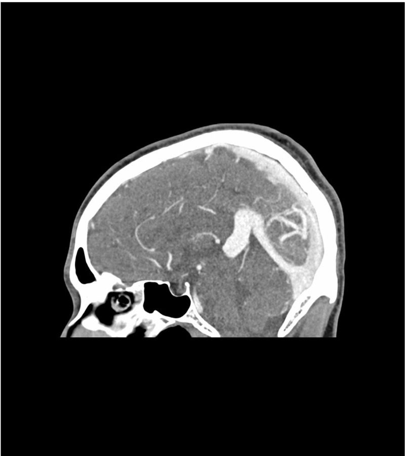
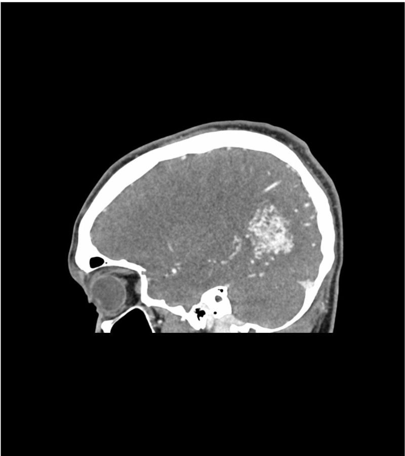
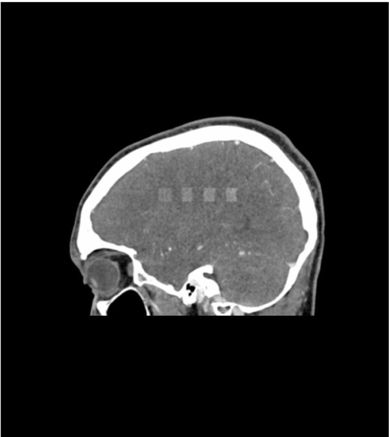
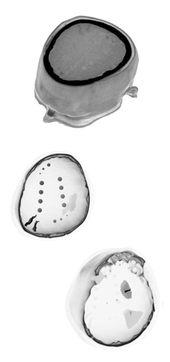
Stroke / Bleed This phantom simulates a head with stroke and bleeding patterns. It covers the vertex to the foramen magnum. Stroke patterns include signs of early infarction (hyperdense middle cerebral artery, disappearing basal ganglia), acute and subacute watershed infarcts, and lacunar infarctions of varying ages. Bleeding patterns include subarachnoid hemorrhage, subdural hemorrhage of varying ages, intraventricular hemorrhage, and intracerebral hemorrhage. The phantom can be used in CT (including CBCT) to evaluate and optimize imaging performance and AI-enabled diagnosis. It is also suited for training purposes. The phantom provides a detailed and realistic simulation of common brain pathologies, soft and bone tissues. Air voids are filled with a cellulose-polymer composite of approx. -160 HU. |
 |
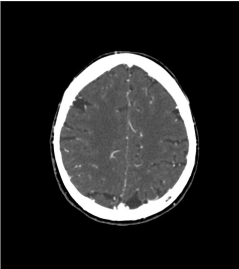
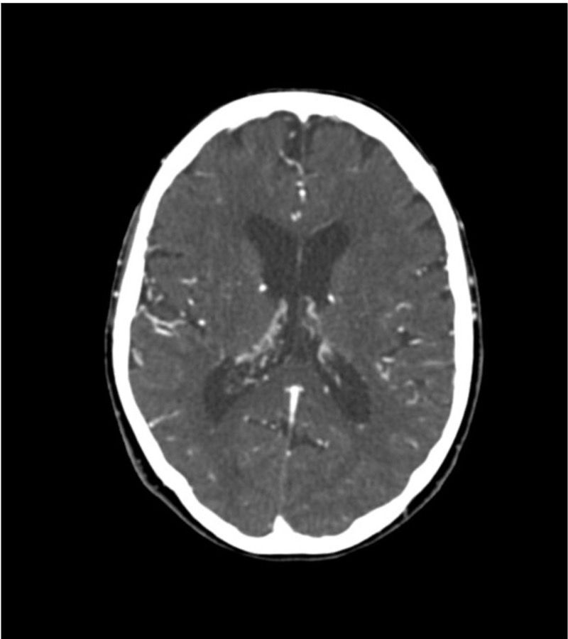
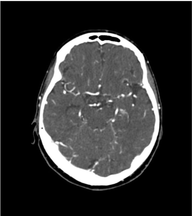
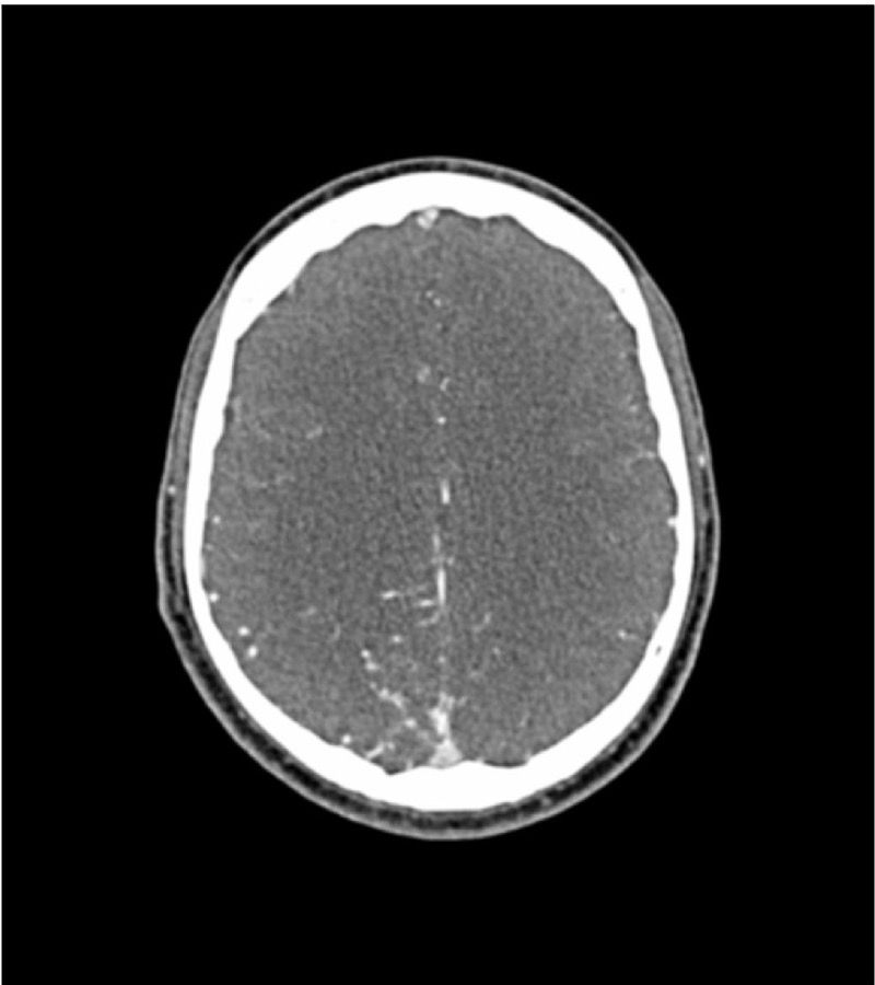
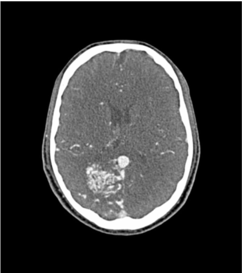
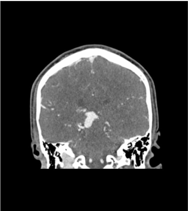
Diagnostic features |
|
|
|
Stroke
Hemorrhage
|
|
|
||||||||||||||||||||||||||||||||||||