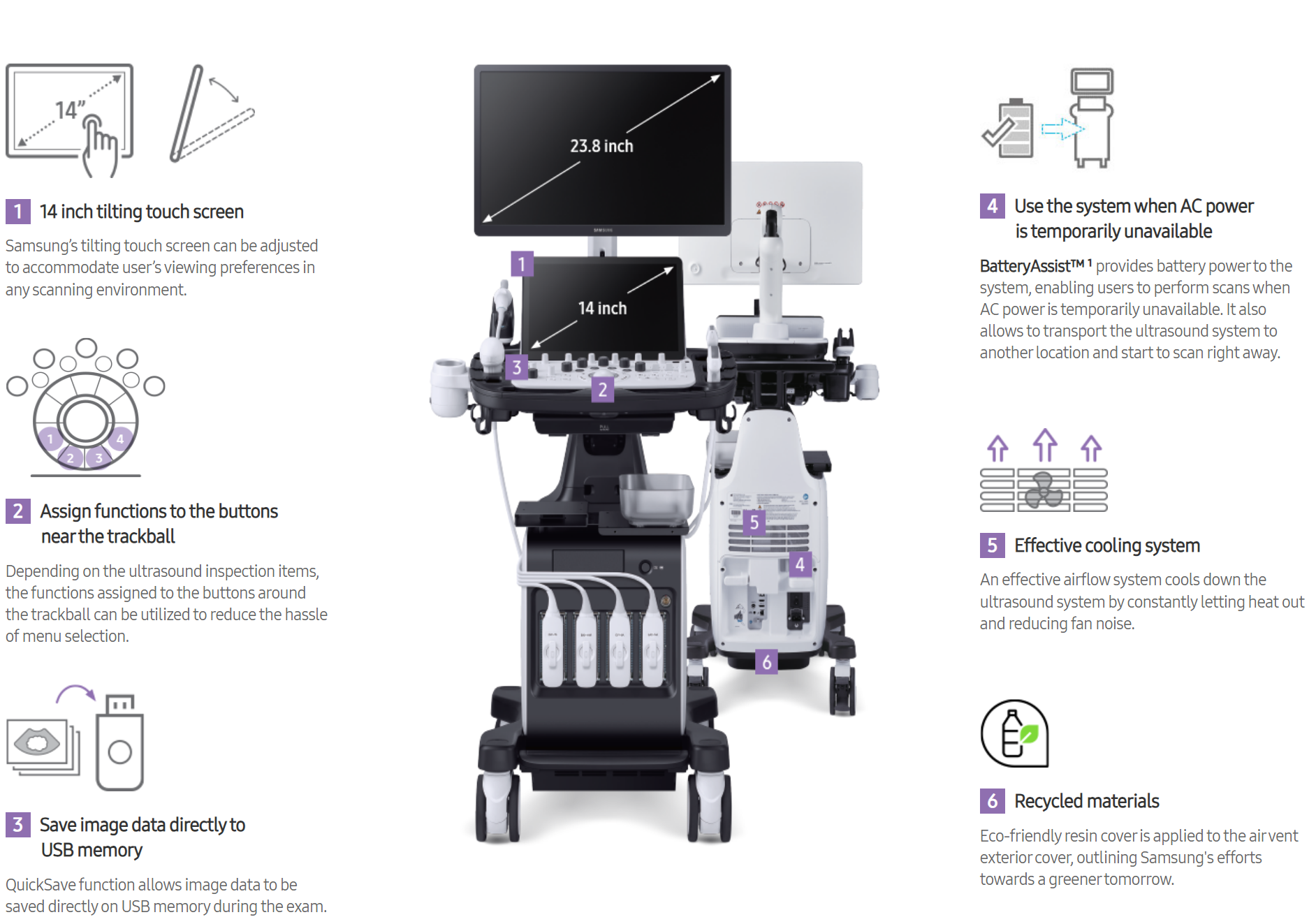
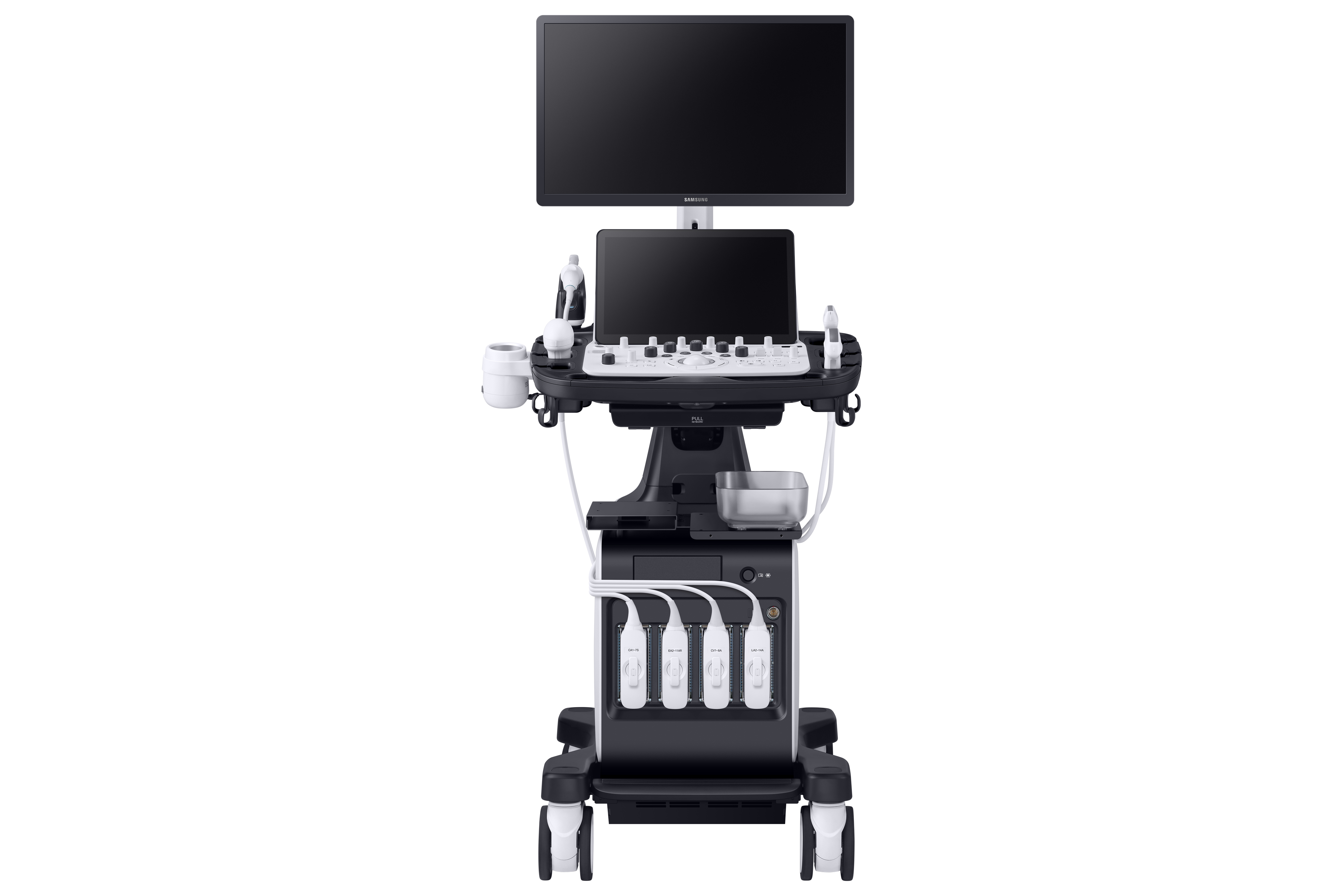
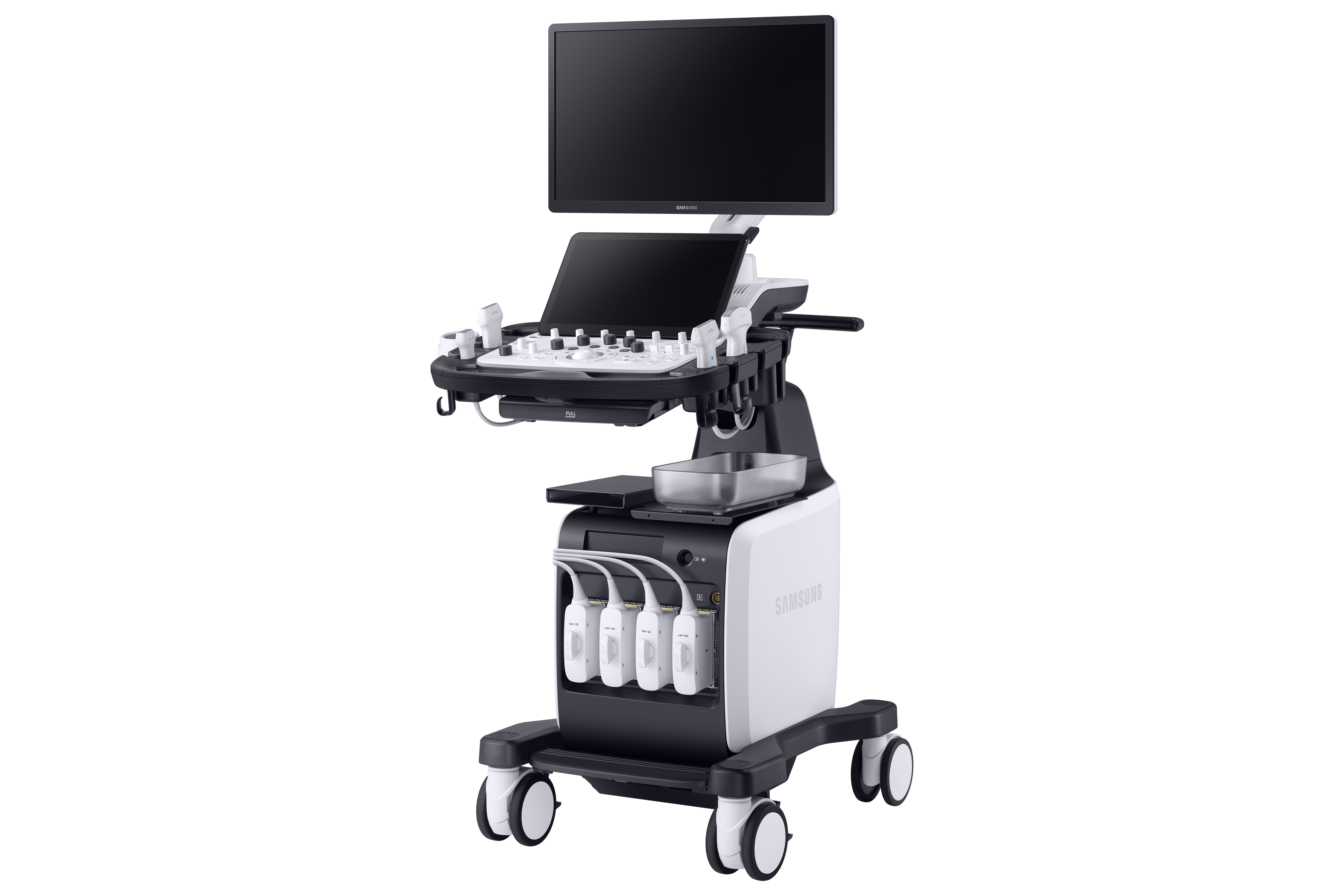
Unifying intelligence and performance
Built to deliver comfort to both healthcare professionals and patients,the V8 premium ultrasound system enhances workflow and patient throughput in women's healthcare. Powered by Samsung's Crystal Architecture™ and Intelligent Assist features, V8 helps streamline processes and boost confidence even in complex women's exams,as well as help communicate results easily with patients.
Redefined imaging technologies powered by Crystal Architecture™
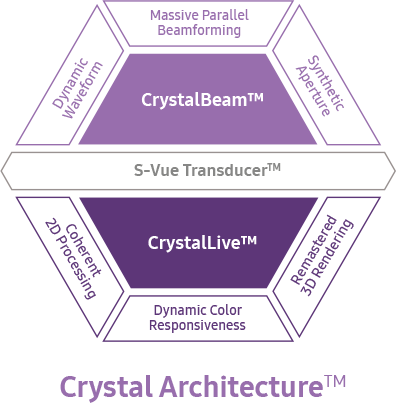 |
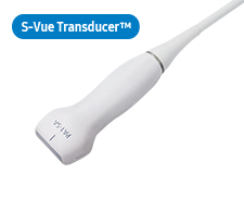
Crystal Architecture™, an imaging architecture that combines CrystalBeam™ and CrystalLive™, based upon S-Vue Transducer™,provides a crystal clear image. CrystalBeam™ is a new beamforming technology beneficial in delivering high-quality image resolution and increased uniformity of images. CrystalLive™ is Samsung’s up-todate ultrasound imaging engine with enhanced 2D image processing,3D rendering and color signal processing, to offer outstanding image performance and efficient workflow during complex cases. |
![]()
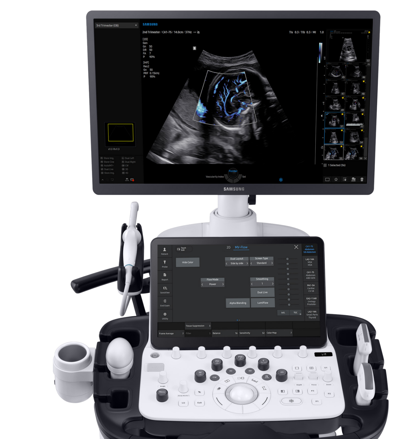
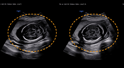
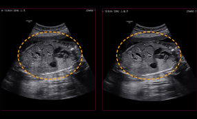
Exquisite imaging qualityfor reliability and confidence
Gain insight into the problem based on exceptional image performance powered by Samsung’s core imaging engine, Crystal Architecture™. The premium imaging engine combines the benefits of enhanced 2D image processing, realistic 3D rendering and detailed expression of color signal processing.
![]()
Enhance hidden structures in shadowed regions
ShadowHDR™ selectively applies high-frequency and low-frequency of the ultrasound to identify shadow areas such as fetal head or spine where attenuation occurs.
Reduce noise to improve 2D image quality
ClearVision The noise reduction filter enhances the edge contrast and creates sharp 2D images for optimal diagnostic performance. In addition, ClearVision provides application-specific optimization and advanced temporal resolution in live scan mode.
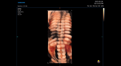
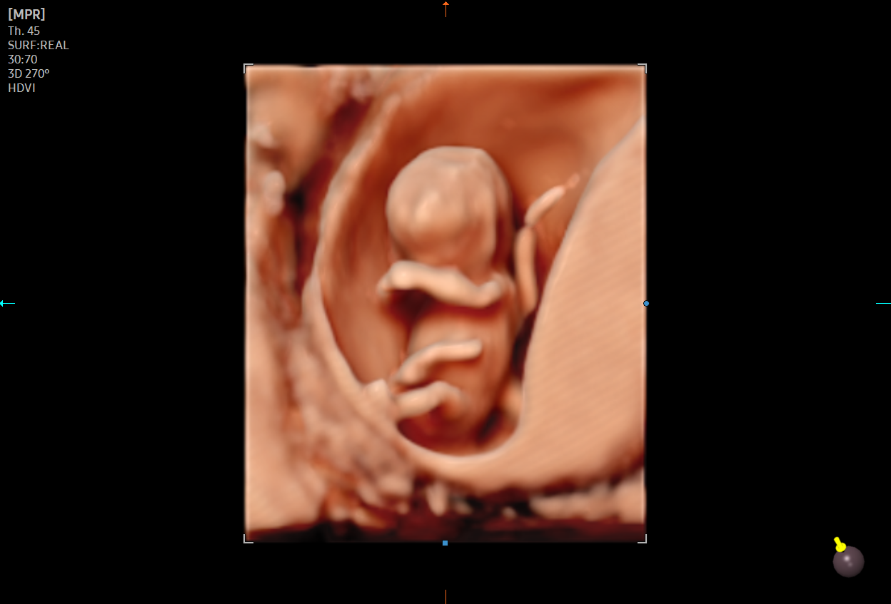
High definitionvolume imaging
HDVI™ is a volume filtering technology that improves visualization of edges and small structures in volume data. Upgraded marginal expression and image saturation expresses the very details from angle to shadow of the fetus.

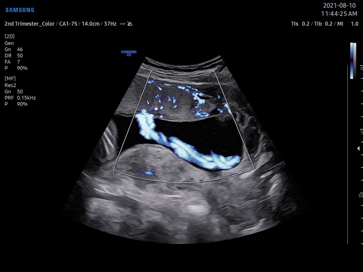
Visualize slow flow in microvascular structures
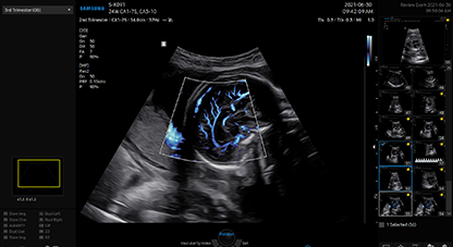
MV-Flow™ offers an advanced color imaging for visualizing slow flow of microvascularized structures. High frame rates and advanced filtering enable MV-Flow™ to provide a detailed view of blood flow in relation to surrounding tissue or pathology with enhanced spatial resolution.
Show blood flow in vessels in a 3D like display
LumiFlow™ is a function that visualizes blood flow in three dimensional-like to help understand the structure of blood flow and small vessels intuitively.
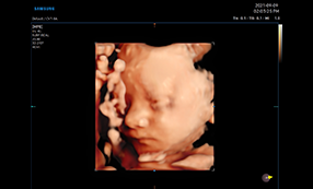
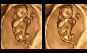
Express 3D anatomyin detailed andrealistic view
RealisticVue™ displays high resolution 3D anatomy with detailed expression and realistic depth perception. User selectable light source direction creates intricately graduated shadows for better defined anatomical structures.
Visualize internal and externalstructures by volumerendering
![]()
Intelligent Assist toolsfor efficient examination
Simplify operations with built-in Intelligent Assist features specialized for obstetrics and gynecology.
V8 supports healthcare professionals with the time-saving features they need in today’s busy working environment.
The system is equipped with a range of tools that help accurately diagnose issues and achieve greater throughput.
For instance, ViewAssist™’s amazing features automatically perform measurements and annotations
with a simple click of a button, thereby reducing repetitive tasks for healthcare professionals.
An automated classification and annotation of the images
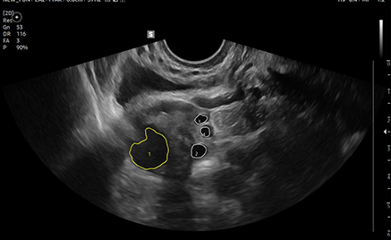
Measure the size of follicles based on 2D
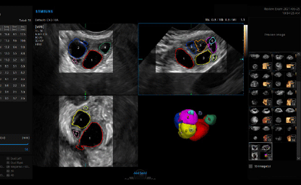
Assess the risk of infertility
Examine patency of the fallopian tube and morphology of uterus and endometrium
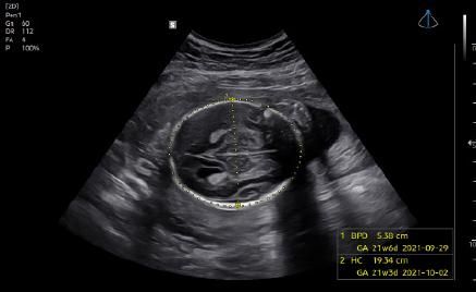
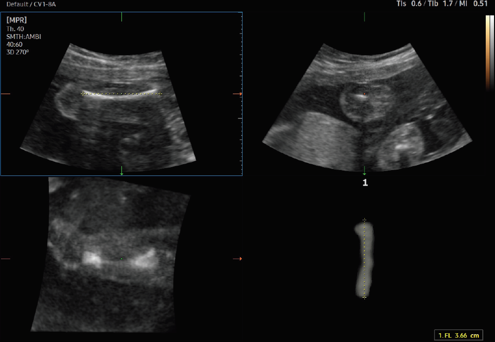
An automated fetal biometry measurement
Estimate fetal weight for checking growth of the fetus
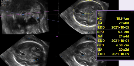
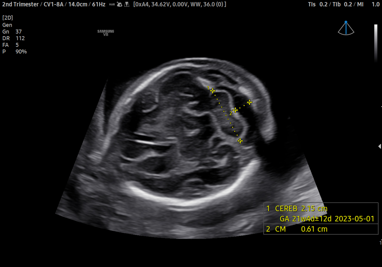
Measure fetal brain in one click
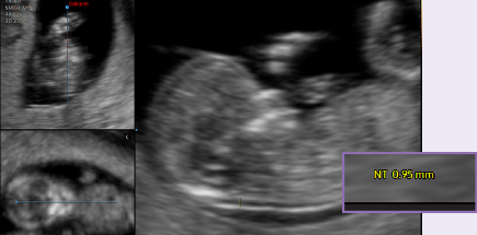
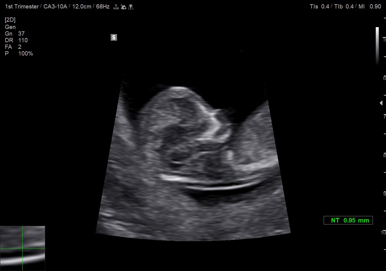
Measure NT using automatic detection of mid-sagittal plane
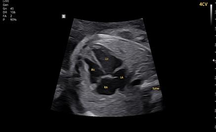
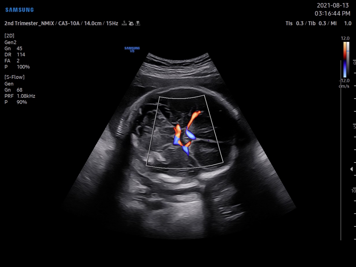
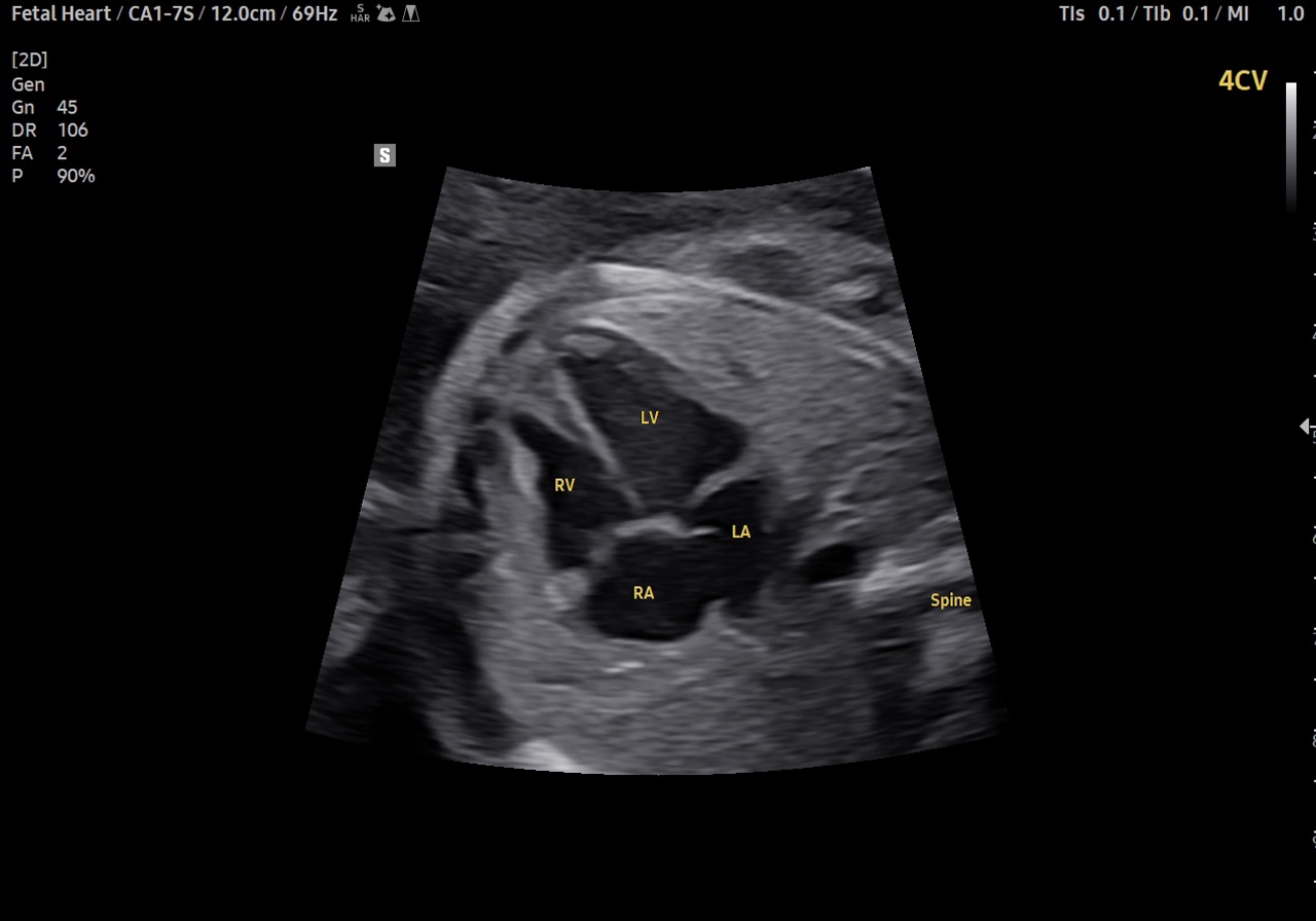
Examine fetal heart including blood flow dynamics
Support in deciding delivery method
Measure stiffness of cervix area for predicting preterm birth
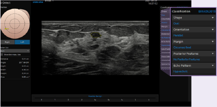
Analyze selected breast lesions and report breast assessment
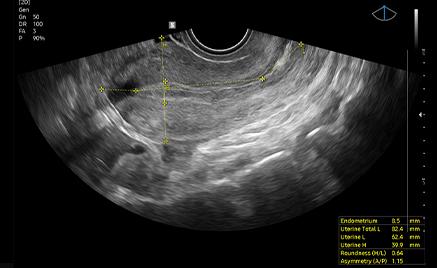
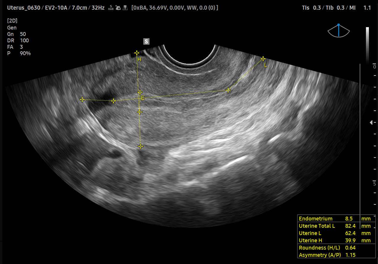
Measure the size and shape of the uterus with AI technology
Classify ovarian tumor
 |
 |
 |
||
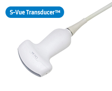
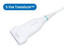
CA1-7S*Application:Abdomen, Obstetrics, Gynecology, Pediatric, Musculoskeletal, Vascular, Urology, Thoracic |
CA3-10AApplication:Abdomen, Obstetrics, Gynecology, Pediatric, Musculoskeletal, Vascular, Urology, Thoracic |
CA4-10M*Application:Abdomen, Vacular, Pediatric |
 |
 |
 |
 |
|
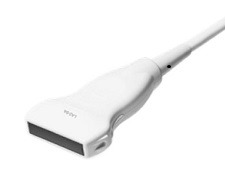
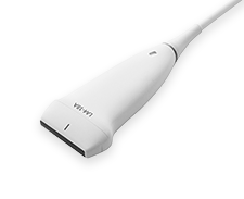
LA2-14AApplication:Small parts, Vascular, Musculoskeletal, Abdomen, Pediatric, Thoracic |
LA2-9AApplication:Small parts, Vascular, Musculoskeletal, Abdomen |
LA2-9SApplication:Small parts, Vascular, Musculoskeletal, Abdomen |
L3-22Application:Musculoskeletal, Small parts, Vascular, Pediatric |
|
 |
 |
|||
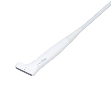
LA4-18A*Application:Small parts, Vascular, Musculoskeletal, Abdomen |
LA3-22AIApplication:Small parts, Vascular, Musculoskeletal, Pediatric, Intraoperative |
 |
 |
|||
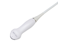
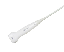
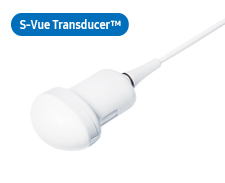
CV1-8AApplication:Abdomen, Obstetrics, Gynecology, Urology |
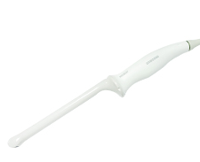
EV2-10A*Application:Obstetrics, Gynecology, Urology |
 |
 |
 |
||
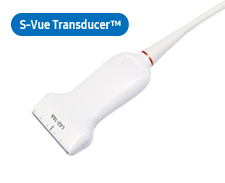
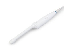
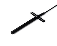
EA2-11AR*Application:Obstetrics, Gynecology, Urology |
EA2-11AV*Application:Obstetrics, Gynecology, Urology |
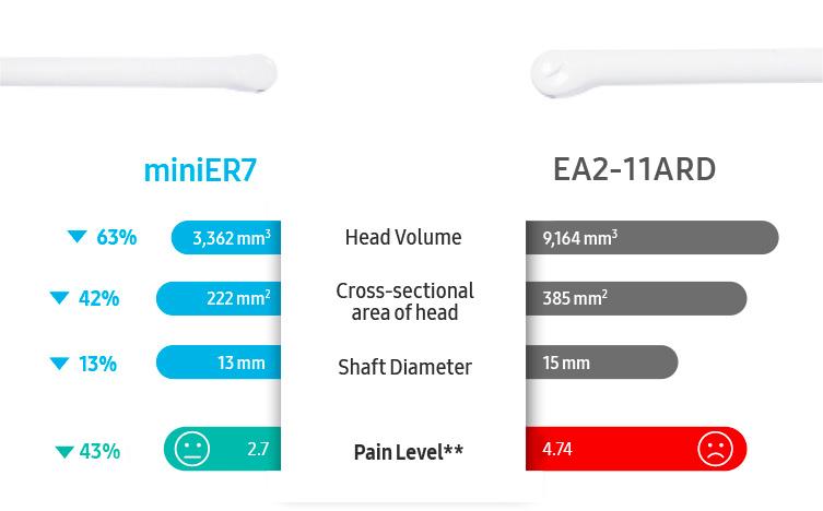
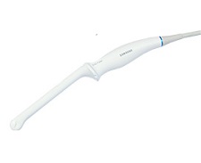
miniER7*Application:Obstetrics, Gynecology, Urology |
 |
 |
 |
||
PA1-5A*Application:Cardiac, Vascular, Abdomen, Pediatric, TCD, Thoracic |
PA4-12BApplication:Abdomen, Cardiac, Pediatric, Vascular, TCD |
PA3-8BApplication:Abdomen, Cardiac, Pediatric, Vascular, TCD |
 |
 |
|||
CW6.0Application:Cardiac |
DP2BApplication:Cardiac |
 |
||||
MMPT3-7Application:Cardiac |
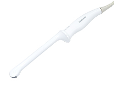
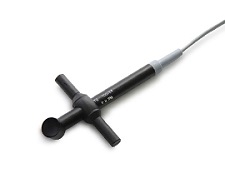
* Ergonomic Transducer
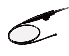
The new convex transducer design with a smooth and slim grip helps users to scan easily and comfortably.
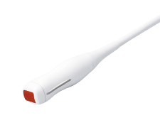
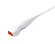
The new endocavity transducer supports natural grip by moving the max width point to a more forward positionand also increased the length of the grip to allow balanced weight distribution.
Ultra Compact Prostate Ultrasound Transducer