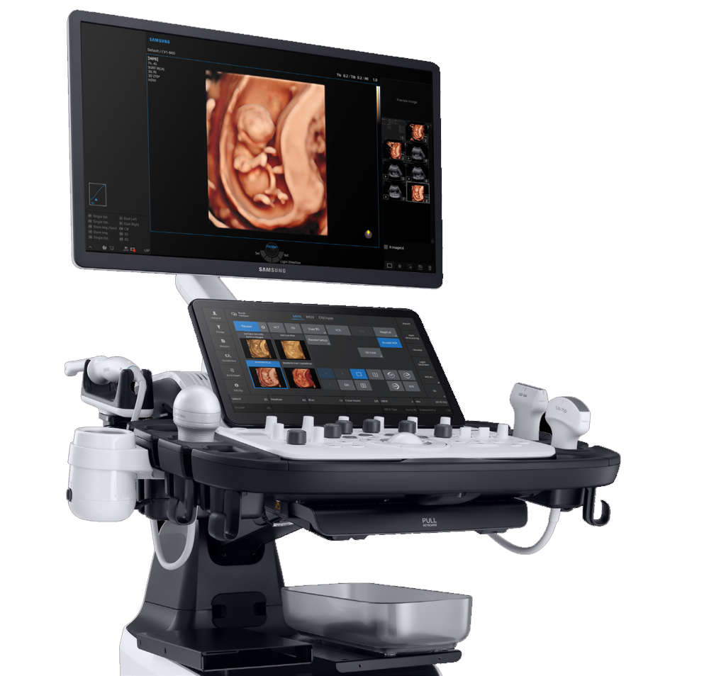
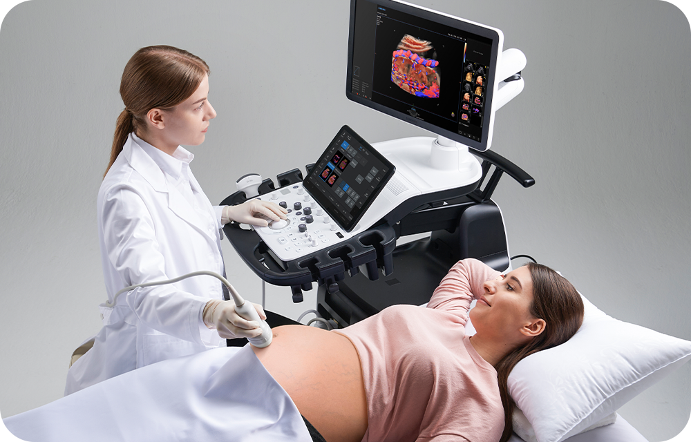
All the key benefits you want
The V7 offers a fascinating performance and gives you the possibility to do what you want with comprehensive tools that feature the latest innovations.
For instance, ViewAssist™'s amazing features automatically perform measurements and annotations with a simple click of a button, thereby reducing repetitive tasks for healthcare professionals.
Rich in features, V7 is fully capable of covering women's health that allows you to explore to the fullest.
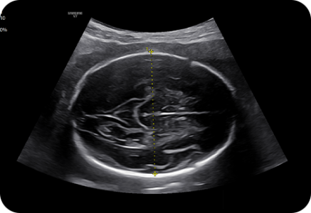
Diagnose diverse and challenging clinical cases
The V7 comes with a variety of tools for diverse and challenging cases.
Healthcare professionals can execute targeted examinations with ease, using the necessary advanced features prepared in the right place.
Furthermore, various sophisticated 2D, 3D, and color imaging features are supported for extraordinary image quality.
2D imaging
 |
|
 |
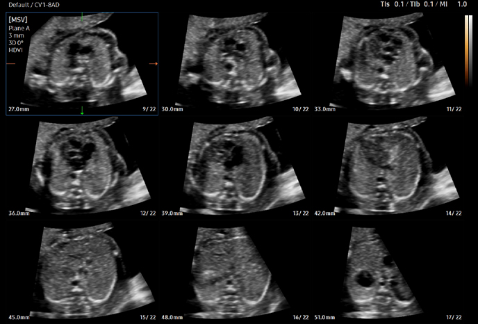
3D imaging
 |
 |
 |
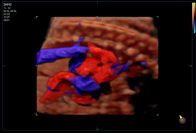
Color imaging
 |
 |
 |
Diagnostic features
 |
 |
 |
 |
 |
 |
 |
 |
![]()
Enriched diagnostic features with accuracy and precision
The V7 system comes with advanced features that assist in precise diagnosis and increasing throughput.
The V7’s variety of features and user-friendly interface aid in significantly improving the healthcare professionals’ daily ultrasound examination experience.
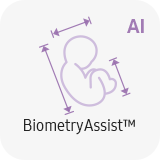
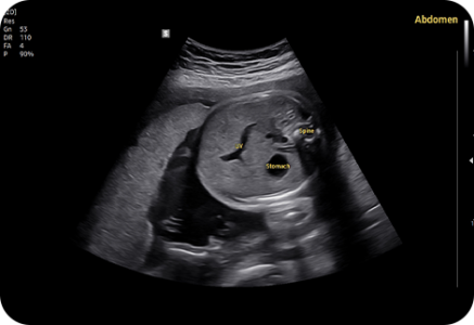
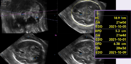
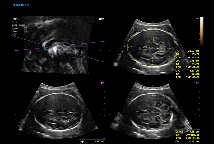
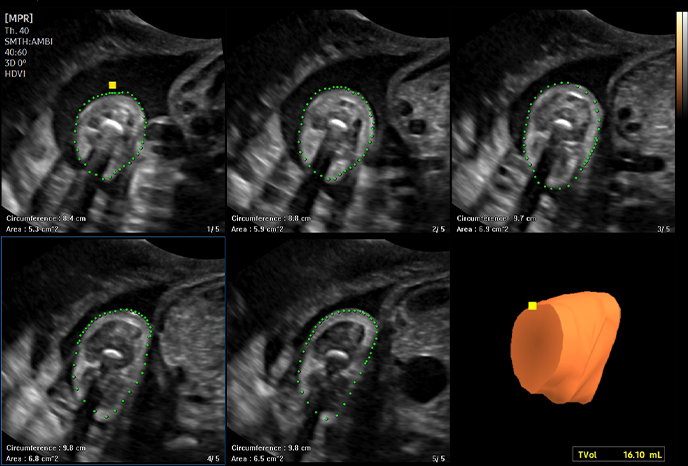
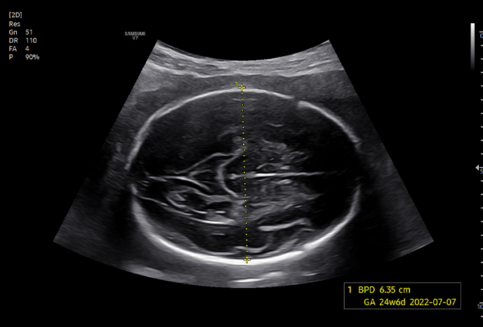
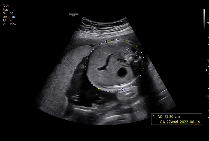
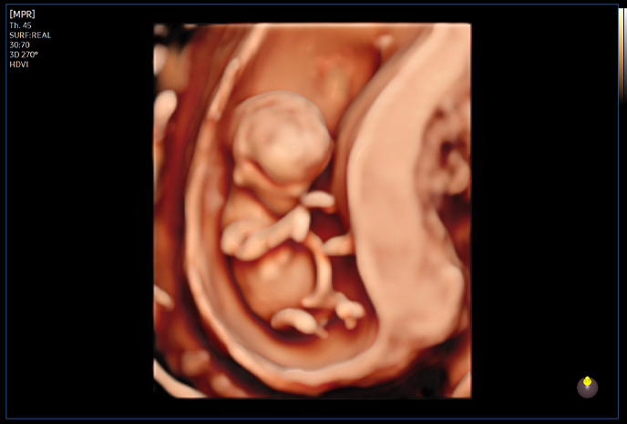
An automated fetal biometry measurement
BiometryAssist™, a feature based on Deep Learning technology, is an automatic technology for biometric measurement. It enables users to measure the fetal growth parameters with one click while maintaining exam consistency.
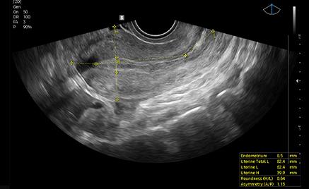
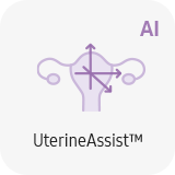
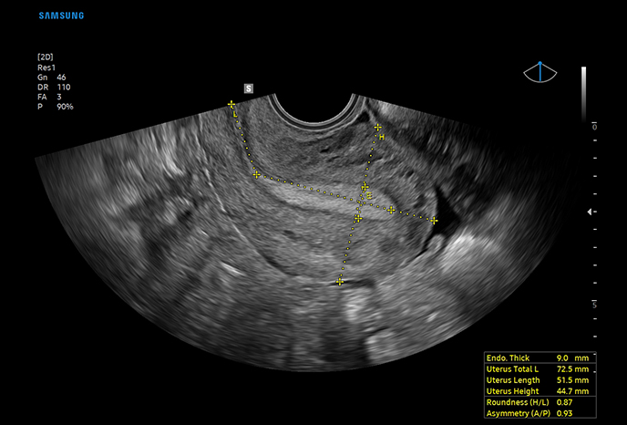
Measure the size and shape of the uterus with AI technology
An automated classification and annotation of the images
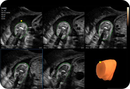
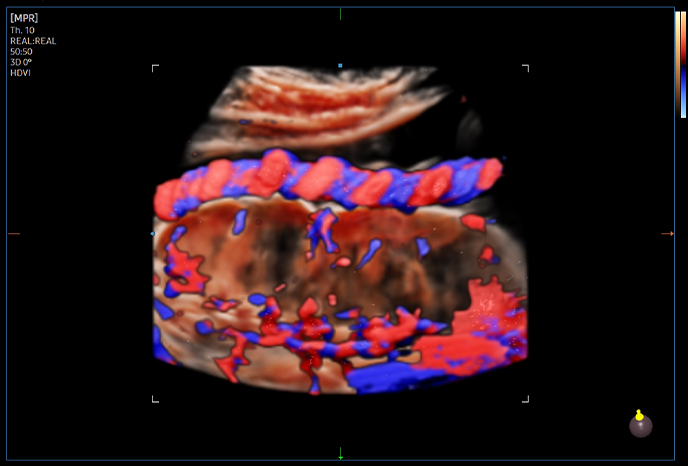
Uterine Contour
Uterine Contour automatically extracts the centerline and thickness of the curved endometrium and provides a coronal view in 3D, flattened by the centerline. In addition, uterine malformation classification are reported according to the ESHRE/ESGE* or ASRM** guideline selection.
* ESHRE/ESGE : The European Society of Human Reproduction and Embryology / The European Society for Gynaecological Endoscopy
** ASRM : The American Society for Reproductive Medicine
Support in deciding delivery method
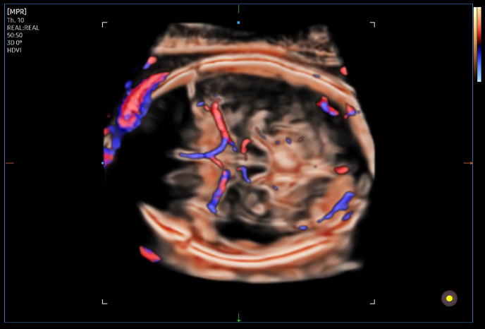
Examine patency of the fallopian tube and morphology of uterus and endometrium
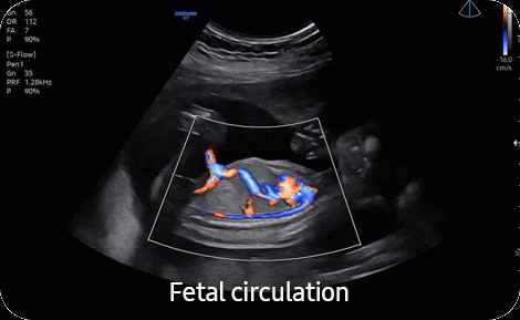
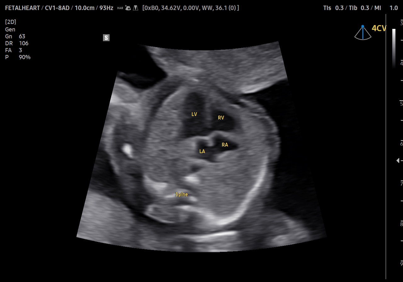
Examine fetal heart including blood flow dynamics
Measure stiffness of cervix area for predicting preterm birth
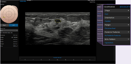
Analyze selected thyroid lesions and report thyroid assessment
S-Detect™ for Thyroid ¹, ⁴ analyzes selected lesions in the thyroid ultrasound study and shows the analysis data, provides standardized reporting based on the ATA, BTA, EU-TIRADS, and K-TIRADS* guidelines; and helps diagnosis with the streamlined workflow.
* ATA: American Thyroid Association
BTA: British Thyroid Association
EU-TIRADS: European Thyroid Imaging Reporting and Data System
K-TIRADS: Korean Thyroid Imaging Reporting and Data System
Estimate fetal weight for checking growth of the fetus
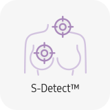
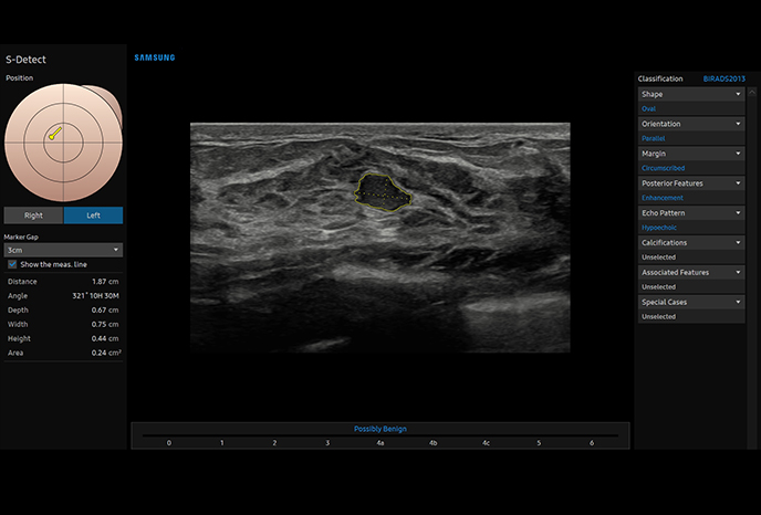
Analyze selected breast lesions and report breast assessment
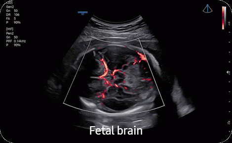
Measure fetal brain in one click
Classify ovarian tumor
Display tissue stiffness in color image
A diagnostic ultrasound technique for imaging elasticity, ElastoScan+™ observes the transformation of the tissue strain by the internal or external forces, and converts relative stiffness into a color image.
asily calculate the strain ratio between two ROIs
E-Strain™ is designed to enable quick and easy calculation of the strain ratio between two regions of interest for day-to-day practice. Simply by setting the two targets, you can receive accurate, consistent results and make informed decisions in many types of diagnostic procedures.
Measure LV MPI and RV MPI semi-automatically
MPI+ is able to semi-automatically measure LV MPI and RV MPI, providing a high reproducibility. After acquiring Inflow/Outflow doppler, RV MPI proceeds alignment by utilizing synchronized signals of the heartrate and valve movement. Through the automatic alignment, it provides ICT, IRT, and RV MPI test results.
Measure the size of follicles based on 2D
Assess the risk of infertility
![]()
![]()
Extraordinary image quality delivers diagnostic confidence
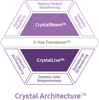 |
Gain insight into complex issues with exceptional image quality and resolution by Samsung’s core imaging engine, Crystal Architecture™. The proprietary technology combines enhanced 2D image processing and detailed color signal processing to optimize and refine the image. |
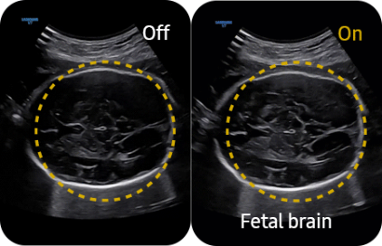
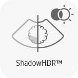
Enhance hidden structures in shadowed regions
ShadowHDR™ selectively applies high-frequency and low-frequency of the ultrasound to identify shadow areas such as fetal head or spine where attenuation occurs.
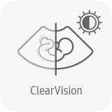
Reduce noise to improve 2D image quality
ClearVision The noise reduction filter enhances the edge contrast and creates sharp 2D images for optimal diagnostic performance. In addition, ClearVision provides application-specific optimization and advanced temporal resolution in live scan mode.
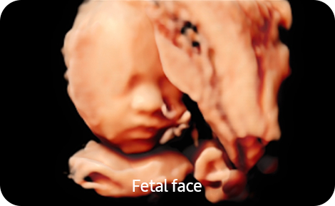
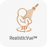
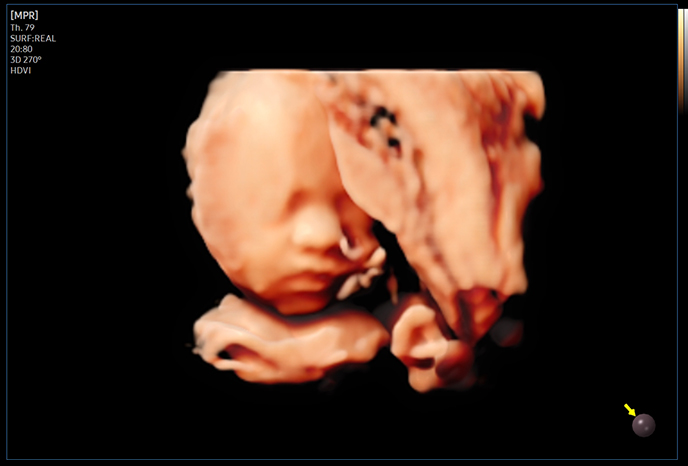
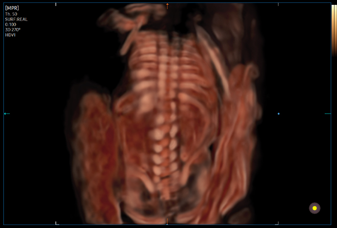
Express 3D anatomyin detailed andrealistic view
RealisticVue™ displays high resolution 3D anatomy with detailed expression and realistic depth perception. User selectable light source direction creates intricately graduated shadows for better defined anatomical structures.
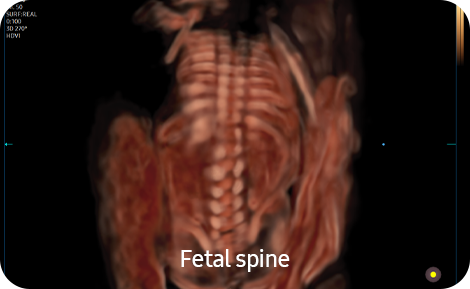
Visualize internal and externalstructures by volumerendering
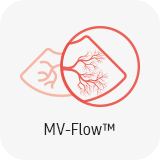
Visualize slow flow in microvascular structures
MV-Flow™ offers an advanced color imaging for visualizing slow flow of microvascularized structures. High frame rates and advanced filtering enable MV-Flow™ to provide a detailed view of blood flow in relation to surrounding tissue or pathology with enhanced spatial resolution.
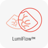
Show blood flow in vessels in a 3D like display
LumiFlow™ is a function that visualizes blood flow in three dimensional-like to help understand the structure of blood flow and small vessels intuitively.
![]()
 |
 |
 |
||
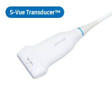
CA1-7SD*Application:Abdomen, Obstetrics, Gynecology, Pediatric, Musculoskeletal, Vascular, Urology, Thoracic |
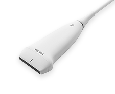
CA3-10AApplication:Abdomen, Obstetrics, Gynecology, Pediatric, Musculoskeletal, Vascular, Urology, Thoracic |
CA4-10M*Application:Abdomen, Vacular, Pediatric |
 |
 |
 |
 |
|
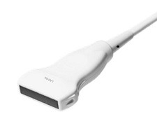
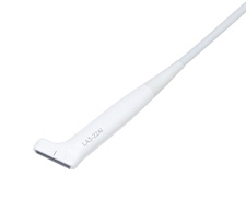
LA2-14AApplication:Small parts, Vascular, Musculoskeletal, Abdomen, Pediatric, Thoracic |
LA2-9AApplication:Small parts, Vascular, Musculoskeletal, Abdomen |
LA2-9S*Application:Small parts, Vascular, Musculoskeletal, Abdomen |
L3-22Application:Musculoskeletal, Small parts, Vascular, Pediatric |
|
 |
 |
|||
LA4-18AD*Application:Small parts, Vascular, Musculoskeletal, Abdomen |
LA3-22AIApplication:Small parts, Vascular, Musculoskeletal, Pediatric, Intraoperative |
 |
 |
|||
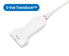
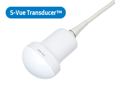
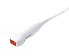
CV1-8ADApplication:Abdomen, Obstetrics, Gynecology, Urology |
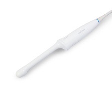
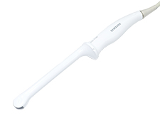
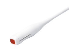
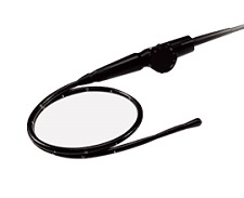
EV2-10A*Application:Obstetrics, Gynecology, Urology |
 |
 |
 |
||
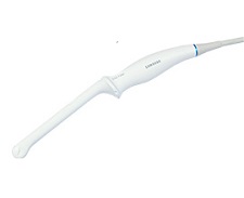
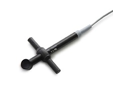
EA2-11ARD*Application:Obstetrics, Gynecology, Urology |
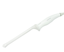
EA2-11AVD*Application:Obstetrics, Gynecology, Urology |
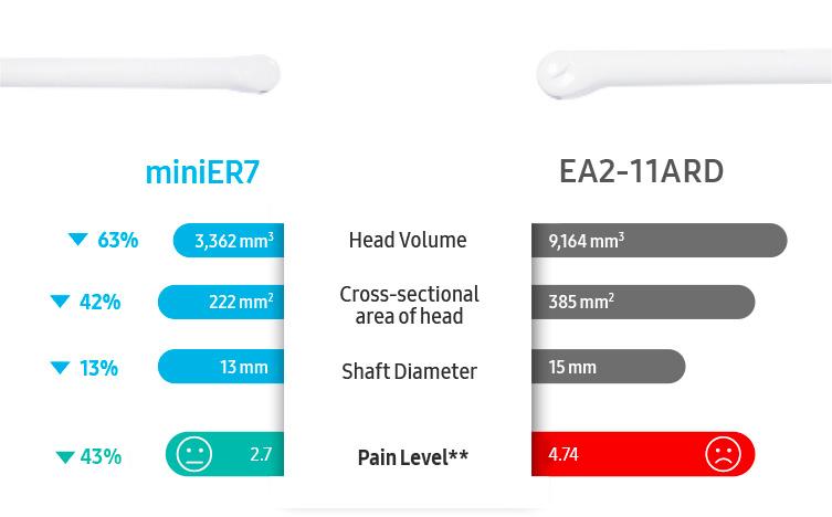
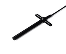
miniER7*Application:Obstetrics, Gynecology, Urology |
 |
 |
 |
||
PA1-5APE*Application:Cardiac, Vascular, Abdomen, Pediatric, TCD, Thoracic |
PA4-12BApplication:Abdomen, Cardiac, Pediatric, Vascular, TCD |
PA3-8BApplication:Abdomen, Cardiac, Pediatric, Vascular, TCD |
 |
 |
|||
CW6.0Application:Cardiac |
DP2BApplication:Cardiac |
 |
||||
MMPT3-7Application:Cardiac |
* Ergonomic Transducer
The new convex transducer design with a smooth and slim grip helps users to scan easily and comfortably.
The new endocavity transducer supports natural grip by moving the max width point to a more forward positionand also increased the length of the grip to allow balanced weight distribution.
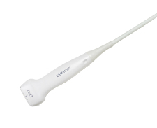
Ultra Compact Prostate Ultrasound Transducer