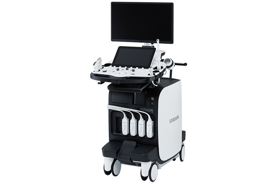
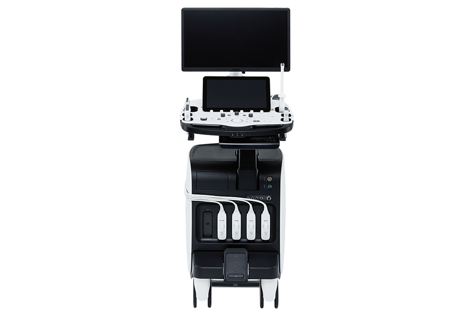
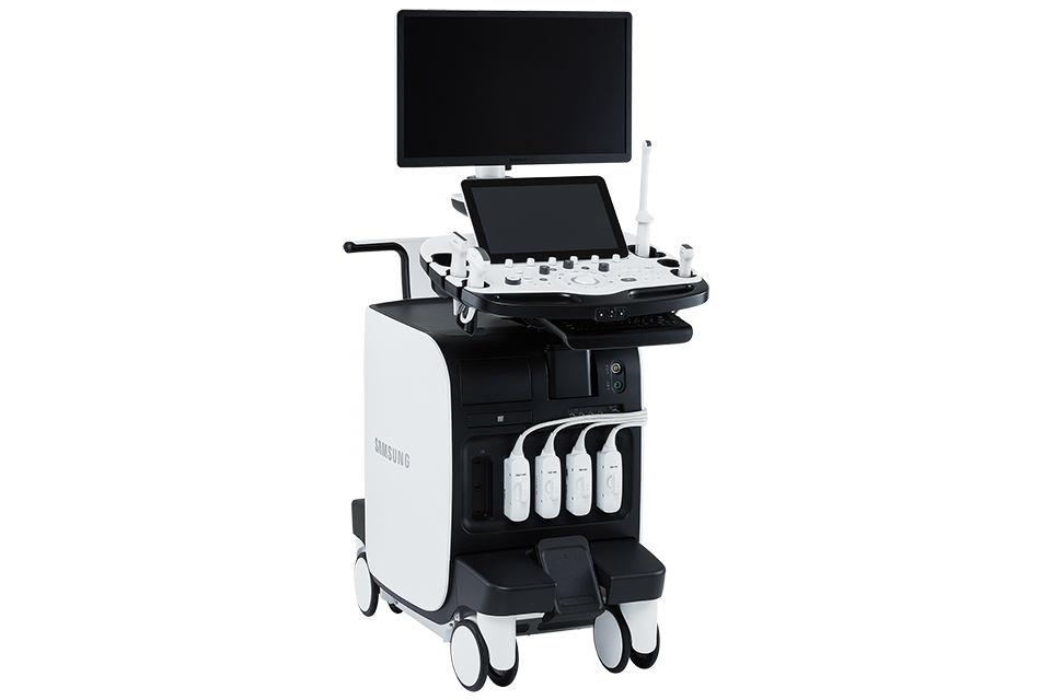
A Revolutionary Change in Advanced Diagnostics
RS85 Prestige has been revolutionized with novel diagnostic features across each application based on the preeminent imaging performance. The advanced intellectual technologies are to help you confirm with confidence for challenging cases, while the easy-to-use system supports your effort involved in the routine scanning.
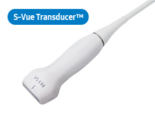
Crystal Architecture™
Crystal Architecture™, an imaging architecture that combines CrystalBeam™ and CrystalPure™, while based upon S-Vue Transducer™, provides crystal clear image.
- CrystalBeam™ is a new beamforming technology beneficial in delivering high-quality image resolution and increased uniformity of images.
Sophisticated 2D & Color Images processed by CrystalPure™
CrystalPure™ imaging engine help you to make more confident diagnoses with fundamental 2D images and enhanced color performance. It also lessens the incidence of clutter and boosts the level of color signal processing.
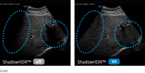
Visualize attenuated shadow area
ShadowHDR™ selectively applies high-frequency and low-frequency of the ultrasound to identify shadow areas where attenuation occurs
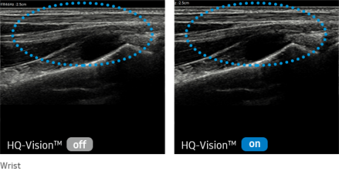
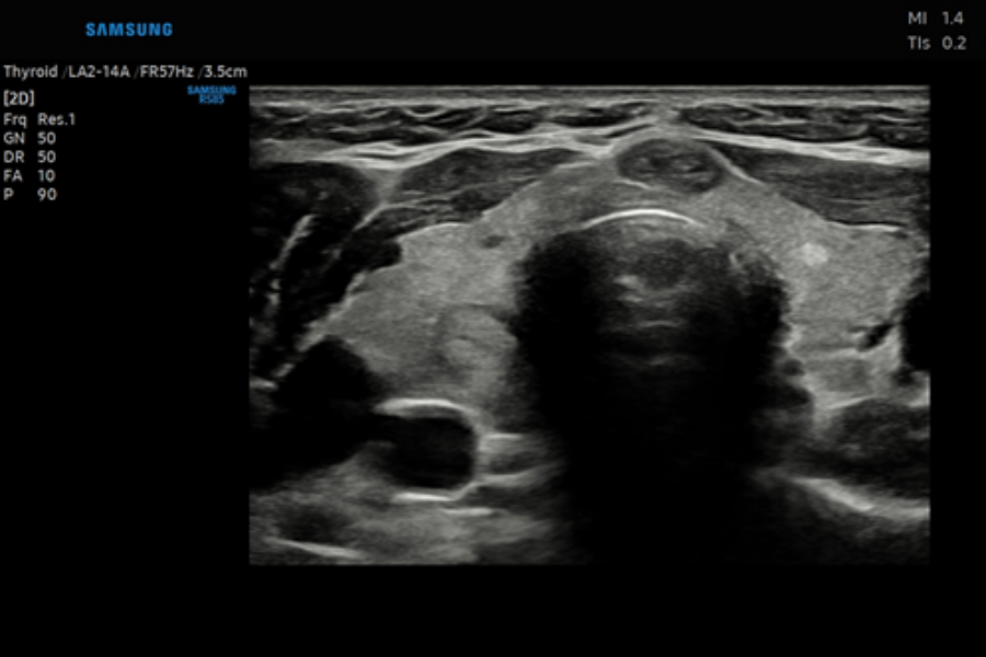
Clarify blurred area to provide clearer images
HQ-Vision™ provides clearer images by mitigating the characteristics of ultrasound images that are slightly blurred than the actual vision.
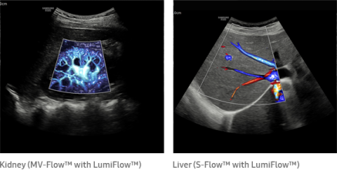
Three dimensional-like visualization of blood flow
LumiFlow™ is a function that visualizes blood flow in three dimensional-like to help understand the structure of blood flow and small vessels intuitively.
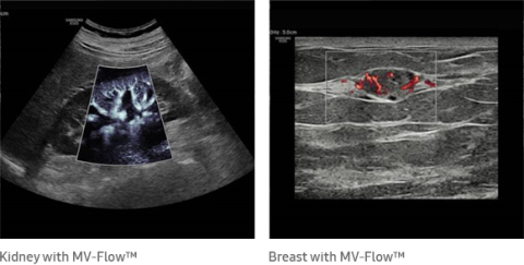
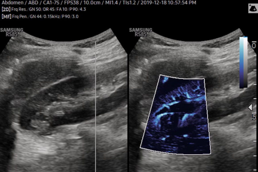
Visualize slow flow microvascularized structures
MV-Flow™ visualizes microcirculatory and slow blood flow to display the intensity of blood flow in color. It is suitable for observation of microcirculatory blood flow and volume of slow blood flow.
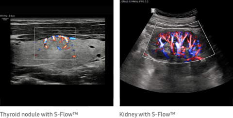
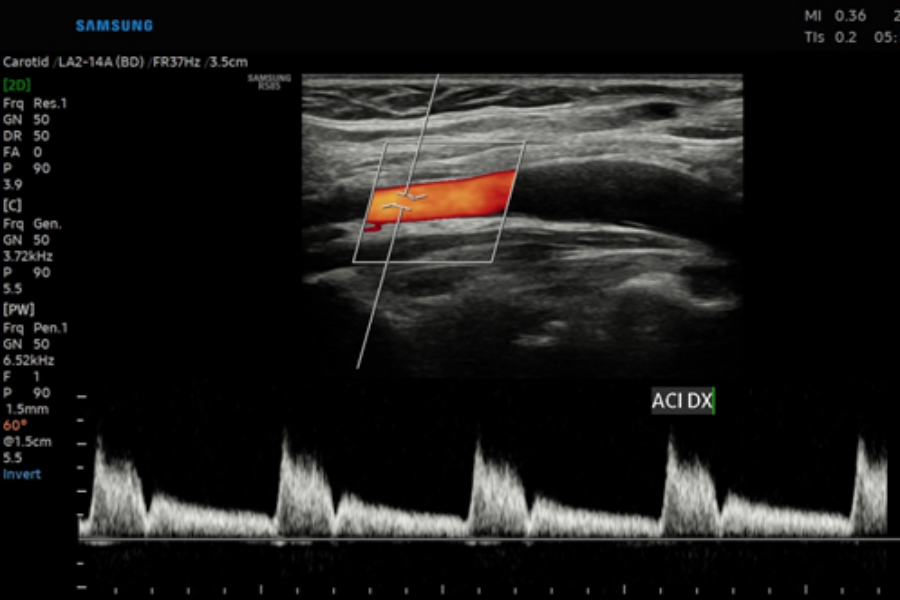
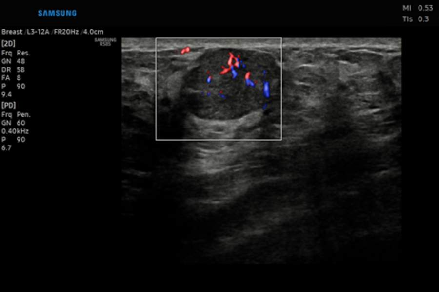
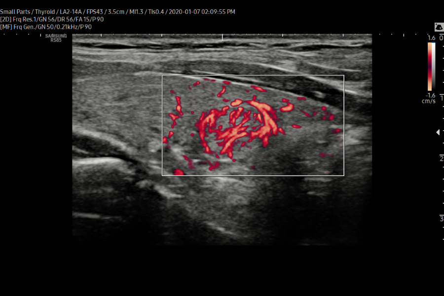
Directional power Doppler to examine peripheral vessels
The function uses directional power Doppler technology, enabling you to examine even the peripheral vessels. It displays information on the intensity and direction of blood flow.
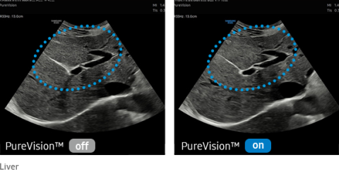
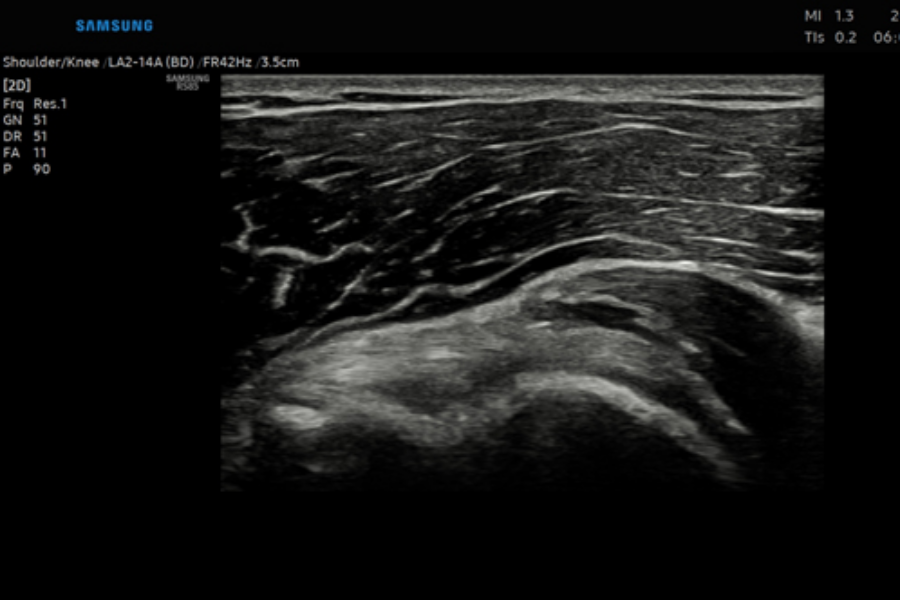
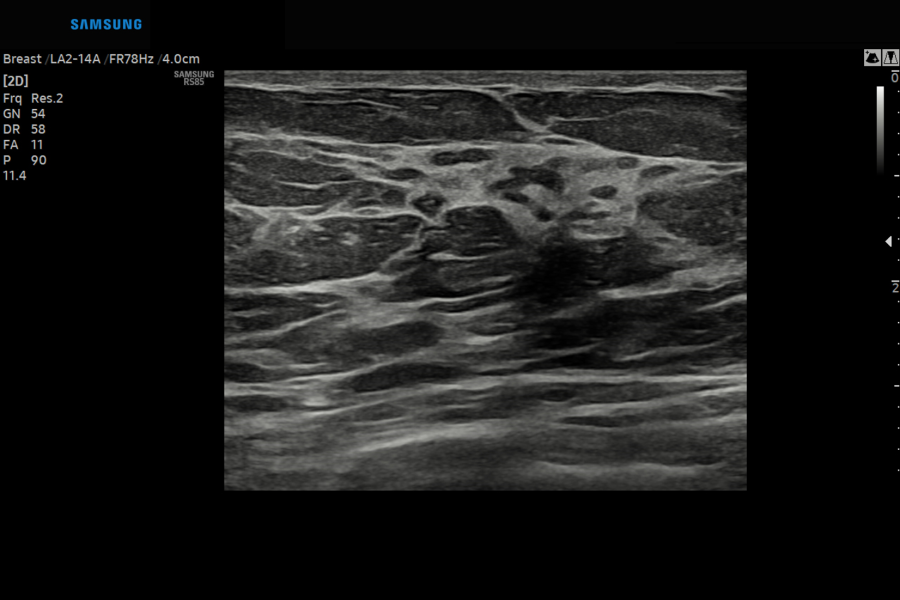
Suppresses speckle noise and enhances edge for dense expression
PureVision™ is an image processing function that outputs with a good uniformity and clear image by performing Speckle Noise Suppression and Edge Enhancement on B-mode.
Advanced Intelligence for Reliable Assessment
Our features enable healthcare professionals navigate and quantify ultrasound propagation in realtime, helping them to visualize and make their assessments with accuracy.
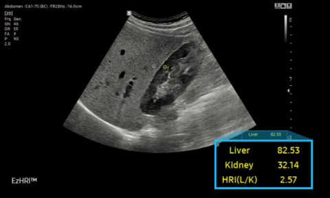
Hepato-renal index (bright comparison) with automated ROI recommendation
HRI (Hepato Renal Index) is an index to quantify steatosis of a liver by comparing echogenicity between liver parenchyma and renal cortex. EzHRITM places 2 ROIs on the liver parenchyma and renal cortex and provides HRI ratio.
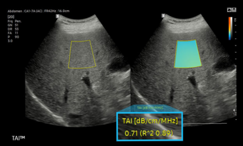
Quantitative measurement of liver fat with ultrasound signal attenuation
TAI™ (Tissue Attenuation Imaging) provides quantitative tissue attenuation measurement to assess steatotic liver changes.
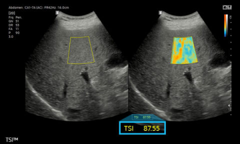
Quantitative measurement of liver fat with with ultrasound signal scatter distribution
TSI™ (Tissue Scatter distribution Imaging) provides quantitative tissue scatter distribution measurement to assess steatotic liver changes.
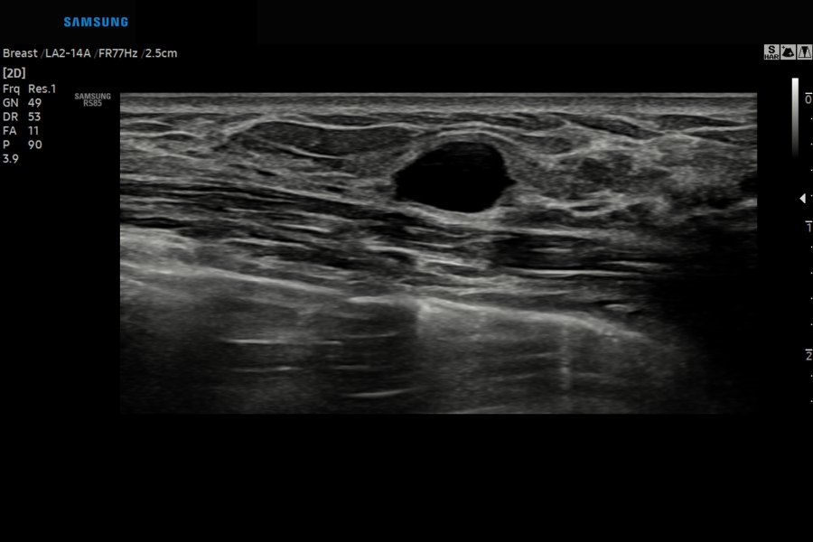
Semi-automated imaging reporting tool for breast assessment
The feature, which analyzes selected lesions in the breast ultrasound study and shows the analysis data, applies BI-RADS ATLAS* (Breast Imaging-Reporting and Data System, Atlas) to provide standardized reporting; and helps diagnosis with the streamlined workflow.
- BI-RADS ATLAS: It is a registered trademark of ACR and all rights reserved by ACR.
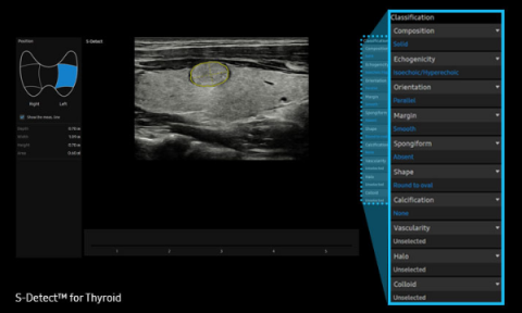
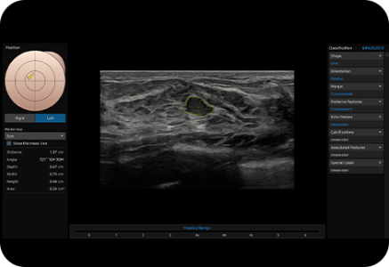
Semi-automated imaging reporting tool for thyroid assessment
The feature, which analyzes selected lesions in the thyroid ultrasound study and shows the analysis data, provides standardized reporting based on the ATA*, BTA*, EU-TIRADS* and K-TIRADS* guidelines and helps diagnosis with the streamlined workflow.
- ATA: American Thyroid Association
- BTA: British Thyroid Association
- EU-TIRADS: European Thyroid Imaging Reporting and Data System
- K-TIRADS: Korean Thyroid Imaging Reporting and Data System
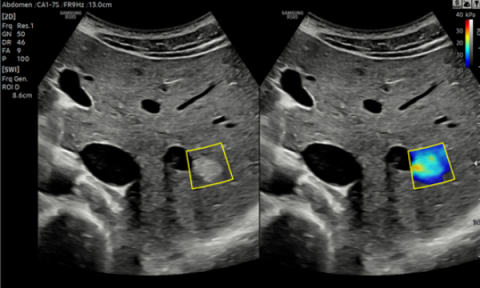
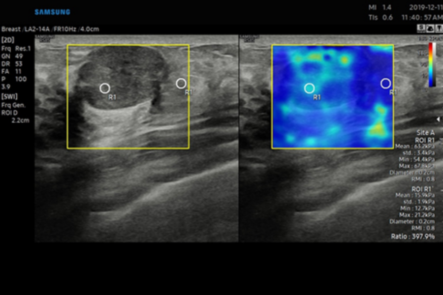
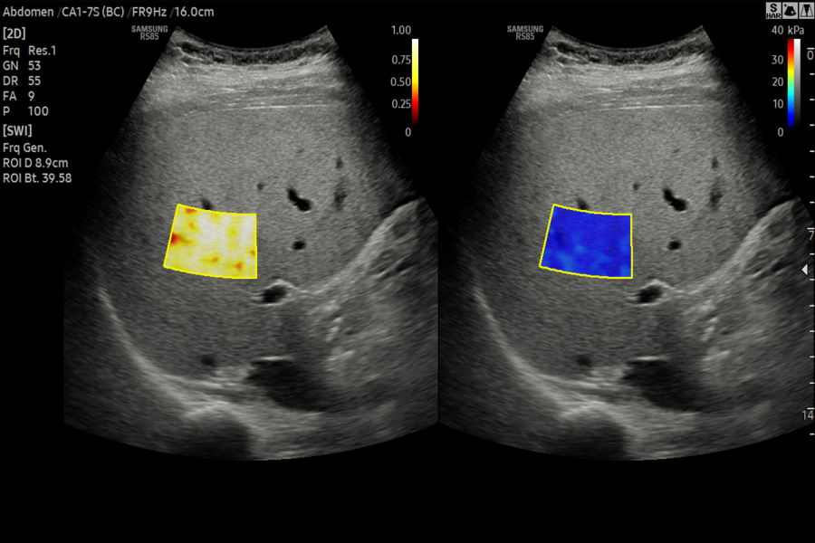
Non-invasive quantification method of tissue stiffness
S-Shearwave Imaging™ allows for non-invasive assessment of the stiffness of tissue/lesions in various applications such as breast, liver, MSK and prostate.The color-coded elastogram, quantitative measurements, dual or single display option, and user-selectable ROI functions are especially useful for the accurate diagnosis of breast and liver diseases.
Precise and Convenient Interventional Solutions
RS85 Prestige provides a broad range of precise fusion, guidance,and dedicated tools to support healthcare professionals strengthen their confidence in operating interventional procedures.
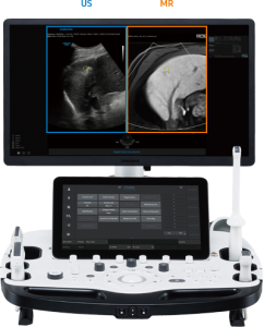
S-Fusion™ for Liver
Intuitive multi-modality fusion imaging with high precision
S-Fusion™ enables simultaneous localization of a lesion using real-time ultrasound in conjunction with other volumetric imaging modalities. Samsung’s Auto Registration helps quickly and precisely fuse the images, increasing efficiency and reducing procedure time. S-Fusion™ enables precise targeting during interventional and other advanced clinical procedures.
Matching Auto
Matching Auto allows automatic initial registration by attaching external markers to the patient’s body before S-Fusion™ exam is processed, thus it helps quick and accurate exam.
Positioning Auto
Positioning Auto helps quick and efficient examination with one-step initial registration between CT/MR and ultrasound images by positioning the transducer in the patient’s pit of the stomach before patient scan.
S-Fusion™ for Prostate
Assists in precise targeting during prostate biopsies
S-Fusion™ for Prostate allows precise targeting during prostate biopsies. Based on 3D models created with MR data sets, S-Fusion™ for Prostate provides biopsy guidance to help safely navigate and target the prostate.
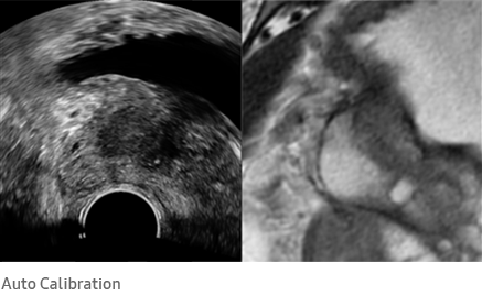
Auto Calibration
S-Fusion™ for Prostate supports a real-time auto calibration function that helps you perform more accurate and reliable procedures.
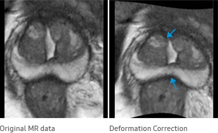
Deformation Correction
Deformation Correction is a feature to improve the accuracy of registration with MR image by correcting deformed prostate shape when transducer is compressed during the procedure and it is useful for targeted biopsy procedure.
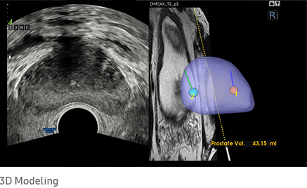
3D Modeling
S-Fusion™ for Prostate allows safe navigation and precise targeting during prostate biopsies based on 3D models created from MR data sets, and also provides a function to report biopsy location.
Enhanced Productivity and Facilitated Workflow
Collaborative solution and streamlined workflow of the RS85 Prestige will support your daily procedures
by reducing keystrokes and by combining multiple actions into one.
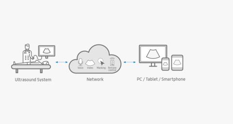
SonoSync™
Real-time image sharing solution
SonoSync™is a real-time image sharing solution that allows collaborative communication and remote controllability for effective collaboration between physicians and sonographers at different locations. Apart from these, SonoSync™ has several other elegant features like marking, invitation, still image sharing, multi-user, and multi-view. SonoSync™ brings telesonography into reality.
SonoSync™ is an image sharing solution, not a diagnostic solution.
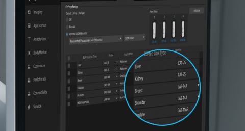
EzPrep™
Automatic transducer setting tool based on the worklist
EzPrep™ is a function that automatically selects the transducer based on the worklist inputted in the ultrasound system and sets the preset of the selected transducer.
RIS Browser
RIS Browser is a function that improves the workflow in the hospital by allowing access to RIS through the browser embedded in the system for the post process without any need to move to the PC after scanning.
 |
 |
 |
 |
 |
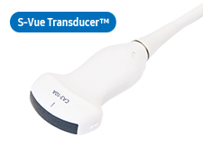
CA1-7AApplication:Abdomen, Obstetrics, Gynecology, Musculoskeletal, Pediatric, Vascular, Urology |
CA3-10AApplication:Abdomen, Obstetrics, Gynecology, Musculoskeletal, Pediatric, Vascular, Urology |
CA2-8AApplication:Abdomen, Obstetrics, Gynecology |
CF4-9Application:Pediatric, Vascular |
CA4-10MApplication:Pediatric, Vascular |
 |
 |
 |
 |
 |
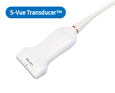
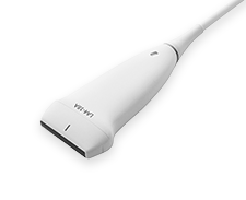
LA2-14AApplication:Small parts, Vascular, Musculoskeletal, Abdomen |
LA2-9AApplication:Small parts, Vascular, Musculoskeletal, Abdomen |
LA2-9SApplication:Small parts, Vascular, Musculoskeletal, Abdomen |
LA3-16AApplication:Small parts, Vascular, Musculoskeletal |
L3-12AApplication:Small parts, Vascular, Musculoskeletal |
 |
 |
 |
 |
 |
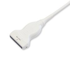
LA4-18AApplication:Small parts, Vascular, Musculoskeletal, Abdomen |
LM4-15BApplication:Small parts, Vascular, Musculoskeletal |
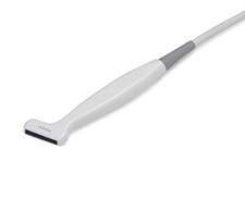
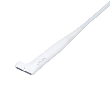
LA3-16AIApplication:Musculoskeletal |
LA3-22AIApplication:Small parts, Vascular, Musculoskeletal, Pediatric, Intraoperative |
LM3-18Application:Small parts, Vascular, Musculoskeletal, abdomen |
 |
 |
|||
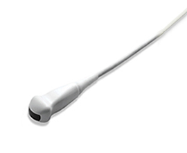
CV1-8AApplication:Abdomen, Obstetrics, Gynecology |
EV3-10BApplication:Obstetrics, Gynecology, Urology |
 |
 |
 |
 |
|
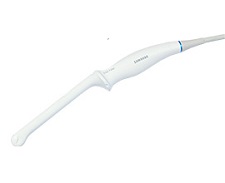
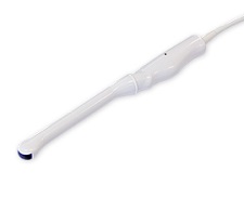
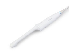
EA2-11AR*Application:Obstetrics, Gynecology, Urology |
EA2-11AV*Application:Obstetrics, Gynecology, Urology |
EA2-11BApplication:Obstetrics, Gynecology, Urology |
EV2-10AApplication:Obstetrics, Gynecology, Urology |
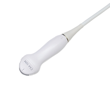
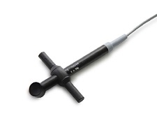
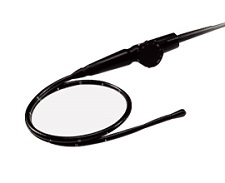
* Ergonomic Transducer (CA1-7S, EA2-11AR, EA2-11AV)
The new convex transducer design with a smooth and slim grip helps users to scan easily and comfortably.
The new endocavity transducer supports natural grip by moving the max width point to a more forward positionand also increased the length of the grip to allow balanced weight distribution.
 |
 |
 |
 |
|
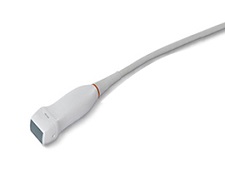
PA1-5AApplication:Cardiac, TCD, Abdomen |
PM1-6AApplication:Cardiac, TCD, Abdomen |
PA3-8BApplication:Cardiac, Pediatric, Abdomen |
PA4-12BApplication:Cardiac, Pediatric |
 |
 |
|||
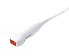
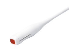
CW6.0Application:Cardiac |
DP2BApplication:Cardiac |
 |
||||
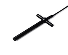
MMPT3-7Application:Cardiac |