Visionary change
Samsung HERA W10 Elite, the premier model of the HERA platform, aims to step up as a visionary leader of Obstetrics and Gynecology applications.
Elegant images, intelligently automated precision features, fast operation, and ergonomics considering the medical environment provide healthcare professionals with diagnostic confidence. The up-to-date system is applied with powerful AI functions to free healthcare professionals from repetitive tasks, in particular, the ViewAssist™ function simplifies the workflow by automatically measuring and annotating numerous views with a simple click of a button.
In addition, the MV-Flow™ 3D function can help detect tiny structures with better sensitivity and resolution, for enhanced clinical decisions. All of these countless advantages are vividly conveyed through the 27-inch OLED monitor.
HERA W10 Elite is our commitment to support the life-long healthcare of women, diligently pursuing new possibilities in ultrasound diagnosis.
Crystal Architecture™
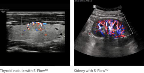
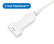
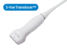
Crystal Architecture™, an imaging architecture that combines CrystalBeam™ and CrystalPure™, while based upon S-Vue Transducer™, is to provide crystal clear image.
- CrystalBeam™ is a new beamforming technology beneficial in delivering high-quality image resolution and increased uniformity of images.
- CrystalPure™ is Samsung’s up-to-date ultrasound imaging engine with enhanced 2D image processing, color signal processing, and advanced intellectual algorithm to offer outstanding image performance and efficient workflow during complex cases.
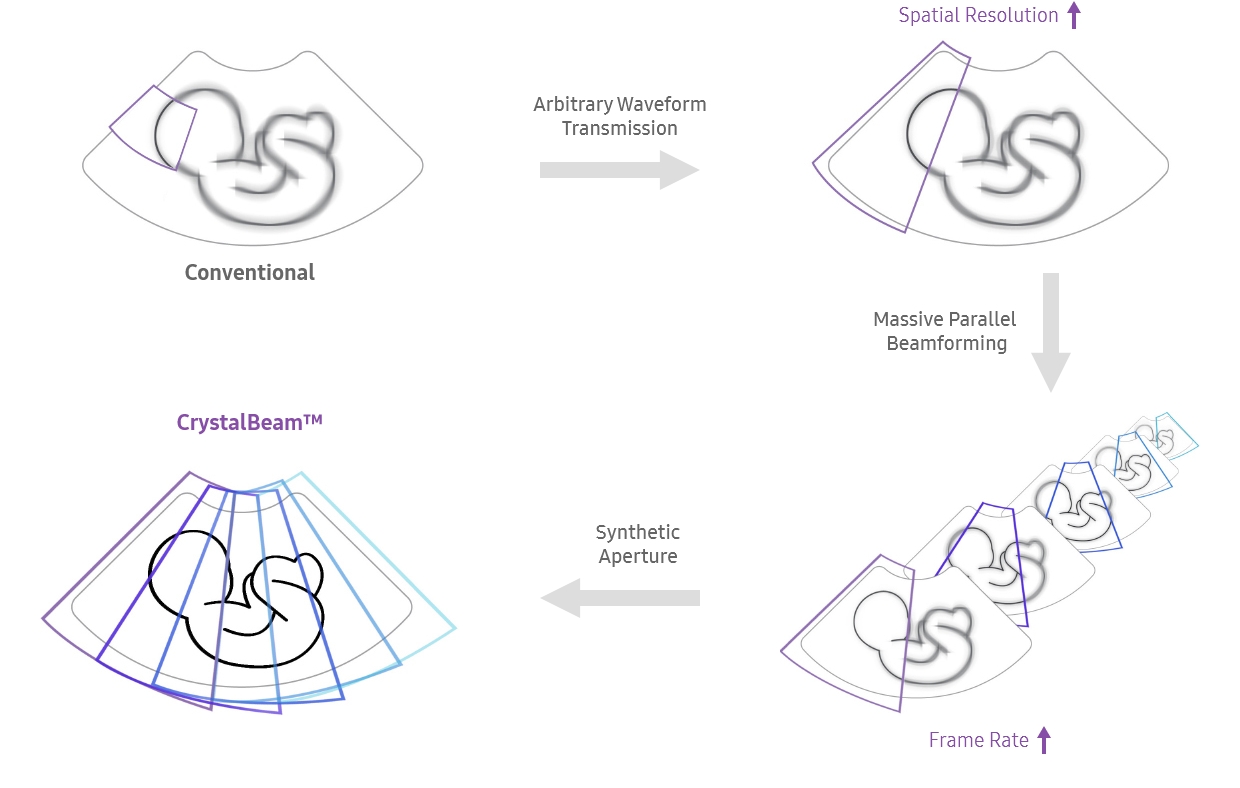
A new beamforming for in-depth image creation
CrystalBeam™ utilizes Arbitrary Waveform Transmission, Massive Parallel Beamforming,
and Synthetic Aperture technologies to produce a faster frame rate and improved image uniformity.
An enriched diagnostic system, excellence in utilization with AI
HERA W10 Elite's expert tools and technologies incorporate the latest artificial intelligence to advance the field. The growth of the fetus and women's health in detailed reports will help you build more confidence and increase the throughput of your diagnosis.
HeartAssist™, a feature based on Deep Learning technology, provides automatic classification of ultrasound image into measurement views required for fetal heart diagnosis and provides measurement results.
ViewAssist™, a feature based on Deep Learning technology, provides automatic classification of the ultrasound images and annotation of the structures to help healthcare professionals in convenient measurement.
* ViewAssist™ fully covers global guidelines from 1st to 3rd trimester.
BiometryAssist™, a feature based on Deep Learning technology, is an automatic technology for biometric measurement. It enables users to measure the fetal growth parameters with one click while maintaining exam consistency.
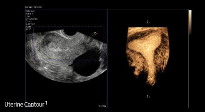
Uterine Contour¹ is a feature to help in identifying uterine malformation. It automatically extracts the centerline and thickness of the curved endometrium and provides a coronal view in 3D, flattened by the centerline. In addition, uterine malformation classification are reported according to the ESHRE/ ESGE or ASRM guideline selection.
* ESHRE/ESGE : The European Society of Human Reproduction and Embryology / The European Society for Gynaecological Endoscopy
* ASRM : The American Society for Reproductive Medicine
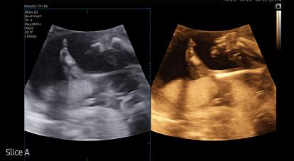
Slice A ¹, is a feature that improves the contrast resolution of A Plane images. By compositing multiple A Plane images, it helps in analyzing tissues or structures that are difficult to see with only 2D images.
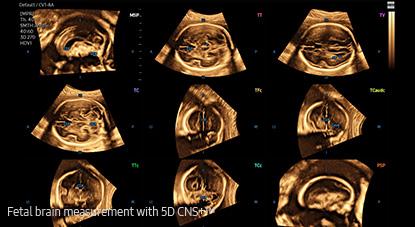
5D CNS+™ ¹ uses intelligent navigation to provide 6 measurements from 3 transverse views of the fetal brain to enhance measurement reproducibility and streamlined workflow.
5D Limb Vol.™ ¹ is a semi-automated tool to quickly and accurately measure upper arm or thigh volumes from 3 simple seed points on a single volume data set. These measurements can then be used to calculate an accurate estimation of fetal weight as well as provide additional information regarding fetal nutritional status.
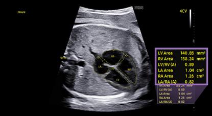
MPI+ is able to semi-automatically measure LV MPI and RV MPI, providing a high reproducibility. After acquiring Inflow/Outflow doppler, RV MPI proceeds alignment by utilizing synchronized signals of the heartrate and valve movement.
Semi-automated imaging reporting tool for breast assessment
The feature, which analyzes selected lesions in the breast ultrasound study and shows the analysis data, applies BI-RADS ATLAS* (Breast Imaging-Reporting and Data System, Atlas) to provide standardized reporting; and helps diagnosis with the streamlined workflow.
- BI-RADS ATLAS: It is a registered trademark of ACR and all rights reserved by ACR.
5D Heart Color™ ¹ The function provides 9 standard planes of the heart by using the fetal STIC data as well as important information about fetal heart development in an easy and accurate way in accordance with the AIUM guideline. In addition, it offers dedicated Preset, Predictive Cursor, Diagnostic Alert, and heart Diastole/Systole timepoints.
5D Follicle™ identifies and measures multiple ovarian follicles in one scan for rapid assessment of follicular size and status during controlled ovarian simulation. This feature uses 3D volume data to help acquire accurate measurement and reduces user variation.
Sophisticated 2D & Color Images processed by CrystalPure™
CrystalPure™ imaging engine help you to make more confident diagnoses with fundamental 2D images and enhanced color performance. It also lessens the incidence of clutter and boosts the level of color signal processing.
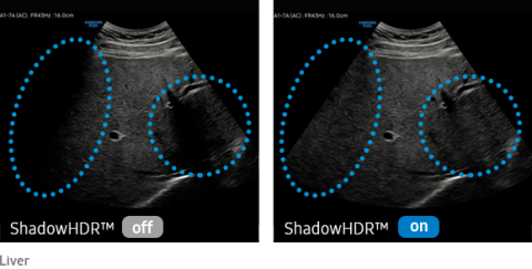
Visualize attenuated shadow area
ShadowHDR™ selectively applies high-frequency and low-frequency of the ultrasound to identify shadow areas where attenuation occurs
ClearVision provides clearer tissue boundaries using the noise reduction filter and generates sharp 2D images. It reduces halo artifact that occurs when the tissue contour is enhanced, and removes noises on the tissue boundaries.
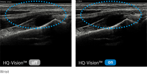
Clarify blurred area to provide clearer images
HQ-Vision™ provides clearer images by mitigating the characteristics of ultrasound images that are slightly blurred than the actual vision.
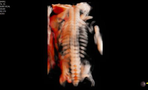
Realistic description of 3D/4D performance
CrystalLive™ in 3D/4D provides users with more realistic and high-resolution images. It outdoes conventional 3D imaging technologies in terms of viewing small parts and lighting effects. In addition, you are able to see 3D anatomy with more realistic depth perception, and can visualize the internal and external structures at once.
CrystalVue™ is an advanced volume rendering technology that enhances visualization of both internal and external structures in a single rendered image using a combination of intensity, gradient and position.
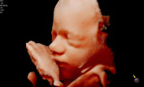
RealisticVue™ ¹ displays high resolution 3D anatomy with exceptional detail and realistic depth perception. User selectable light source direction creates intricately graduated shadows for better defined anatomical structures.
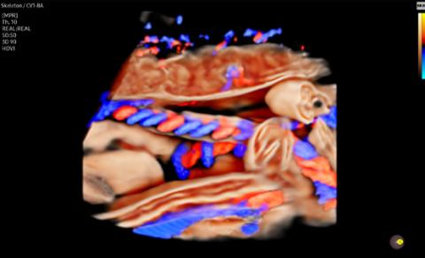
CrystalVue Flow™ ¹ is a volume rendering technology that provides additional information of blood flow morphology based on the CrystalVue™ features that visualizes the internal structures by projecting the 3D data, providing better understanding in the anatomic structures and surrounding vascular vessels.
Advanced vessel description and flow characterization
The color imaging performance has been improved to clearly visualize the hemodynamics of the blood flow. A greater sensitivity resulting from new color signal processing and a realistic 3D visualization of blood flow help understand the microcirculatory blood flows, accurate detection of peripheral blood vessels, and volumes of slow blood flows.
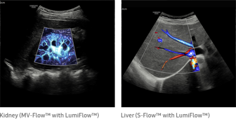
Three dimensional-like visualization of blood flow
LumiFlow™ is a function that visualizes blood flow in three dimensional-like to help understand the structure of blood flow and small vessels intuitively.
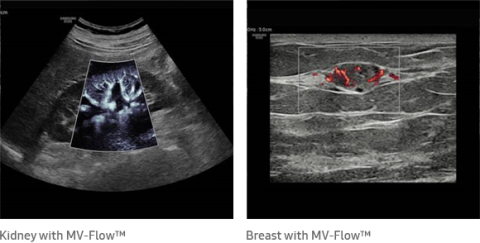
Visualize slow flow microvascularized structures
MV-Flow™ visualizes microcirculatory and slow blood flow to display the intensity of blood flow in color. It is suitable for observation of microcirculatory blood flow and volume of slow blood flow.
Directional power Doppler to examine peripheral vessels
The function uses directional power Doppler technology, enabling you to examine even the peripheral vessels. It displays information on the intensity and direction of blood flow.
 |
 |
 |
 |
|
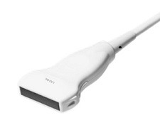
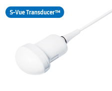
CA1-7AApplication:Abdomen, Obstetrics, Gynecology, Musculoskeletal, Pediatric, Vascular, Urology |
CA3-10AApplication:Abdomen, Obstetrics, Gynecology, Musculoskeletal, Pediatric, Vascular, Urology |
CA2-9AApplication:Abdomen, Obstetrics, Gynecology |
CF4-9Application:Pediatric, Vascular |
 |
 |
 |
||
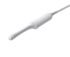
LA2-14AApplication:Small parts, Vascular, Musculoskeletal, Abdomen |
LA2-9AApplication:Small parts, Vascular, Musculoskeletal, Abdomen |
L3-12A
Application:Small parts, Vascular, Musculoskeletal |
 |
 |
 |
 |
|
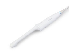
CV1-8AApplication:Abdomen, Obstetrics, Gynecology |
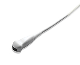
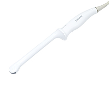
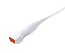
EV3-10BApplication:Obstetrics, Gynecology, Urology |
EV2-10AApplication:Obstetrics, Gynecology, Urology |
EV2-12Application:Obstetrics, Gynecology, Urology |
 |
 |
|||
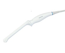
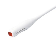
EA2-11AR*Application:Obstetrics, Gynecology, Urology |
EA2-11AV*Application:Obstetrics, Gynecology, Urology |
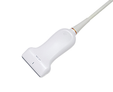
* Ergonomic Transducer (CA1-7S, EA2-11AR, EA2-11AV)
The new convex transducer design with a smooth and slim grip helps users to scan easily and comfortably.
The new endocavity transducer supports natural grip by moving the max width point to a more forward positionand also increased the length of the grip to allow balanced weight distribution.
 |
 |
 |
 |
|
PA1-5AApplication:Cardiac, TCD, Abdomen |
PM1-6AApplication:Cardiac, TCD, Abdomen |
PA3-8BApplication:Cardiac, Pediatric, Abdomen |
PA4-12BApplication:Cardiac, Pediatric |