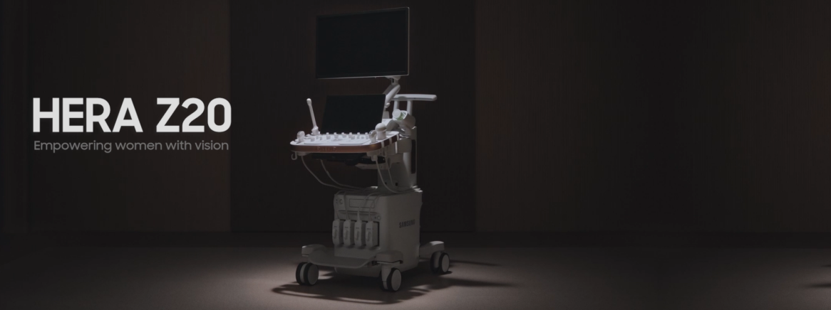

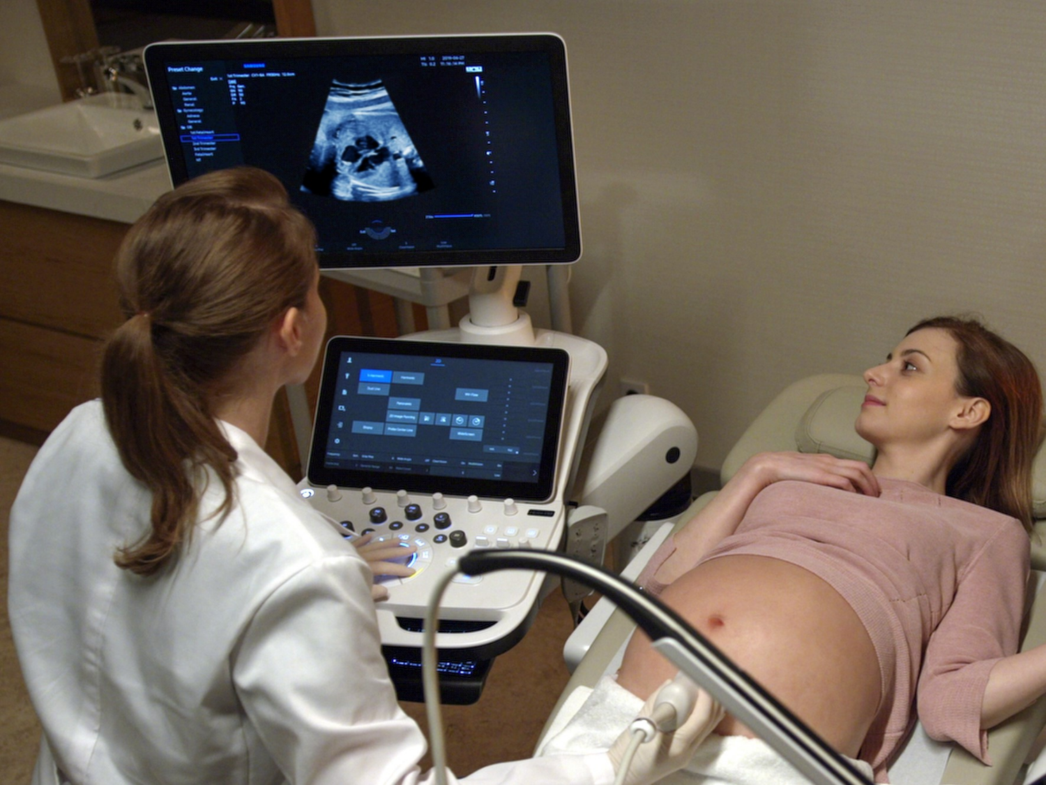
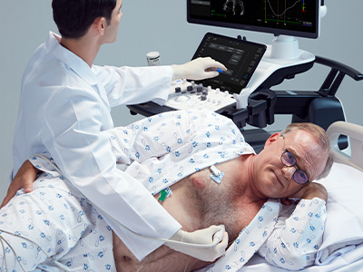
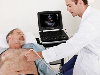
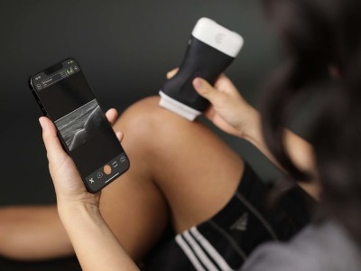
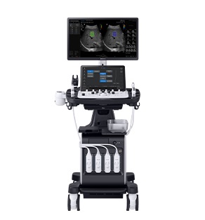
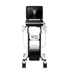
Ultrasound Systems


From our blog
Latest posts from our news room.
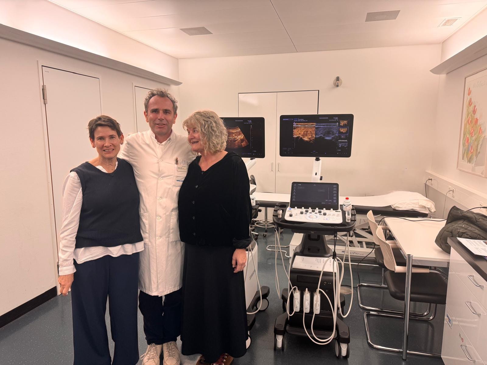
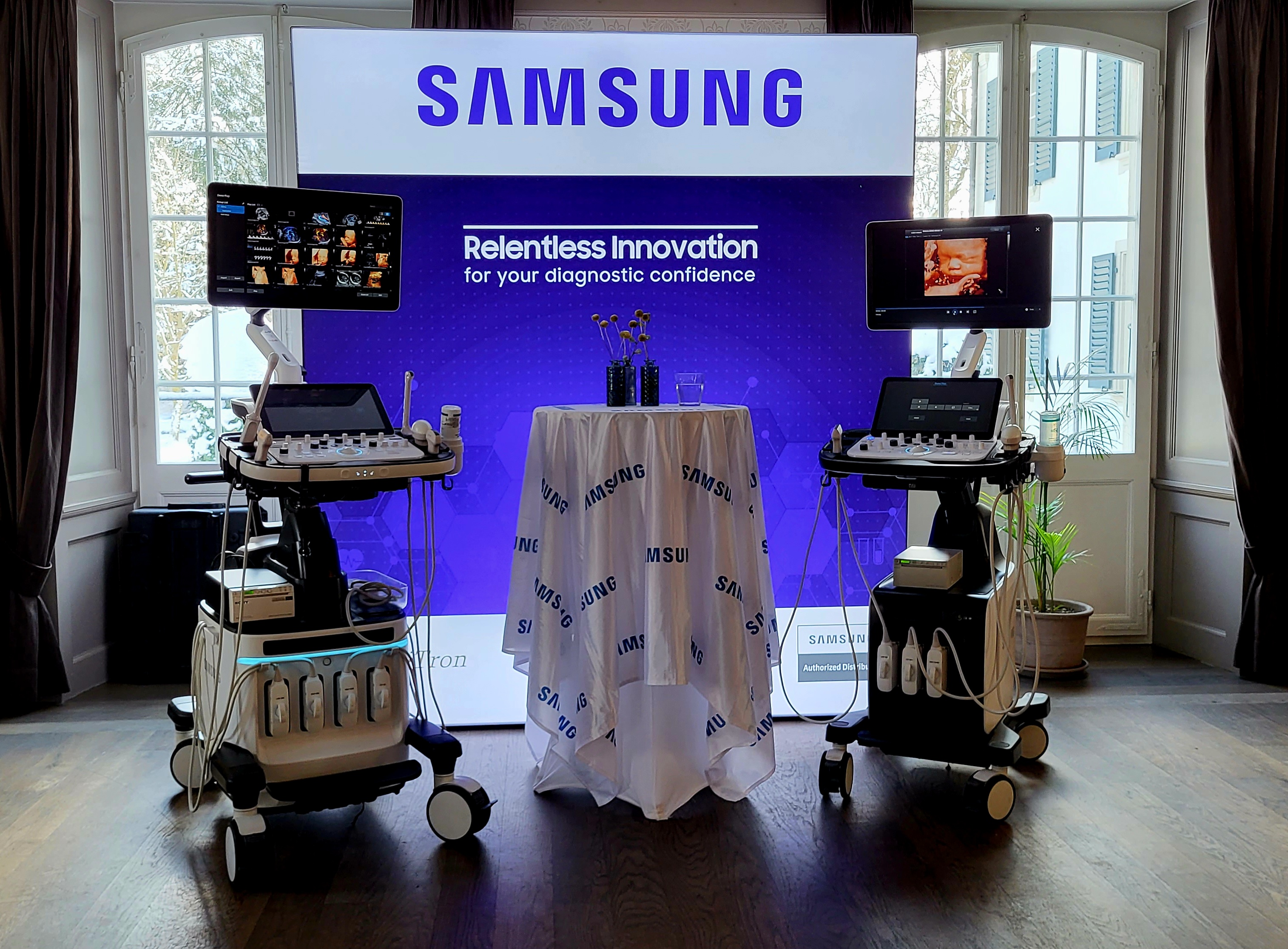
29-01-2025
Professor Andreas Serra, accompanied by Karen and Pia, was presented with two units d...
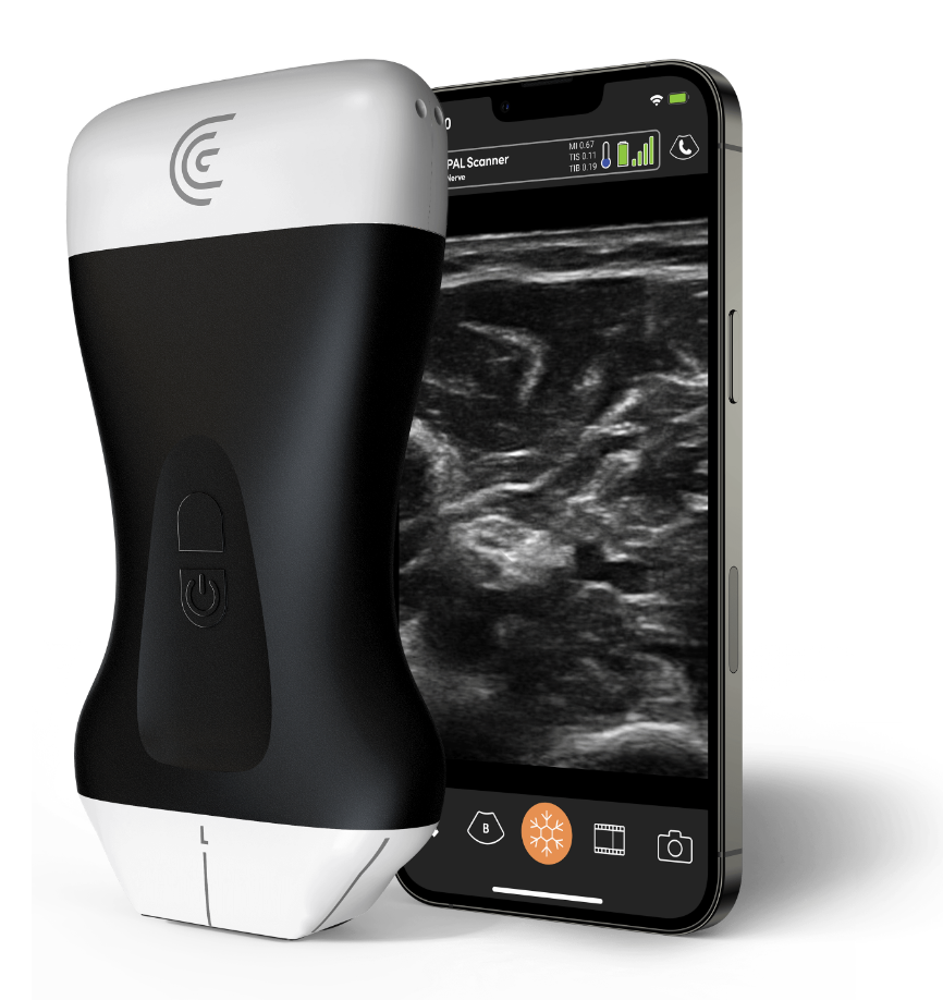
14-03-2024
Another batch of V7s going to our newly acquired customer
The installation was ...
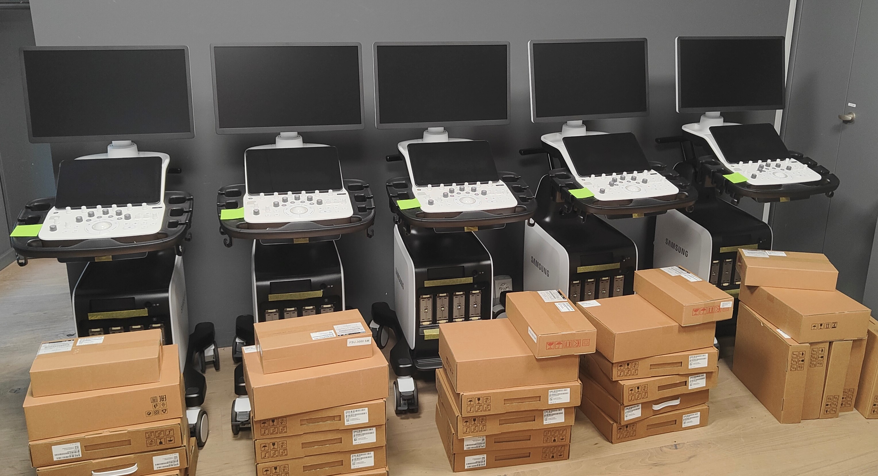
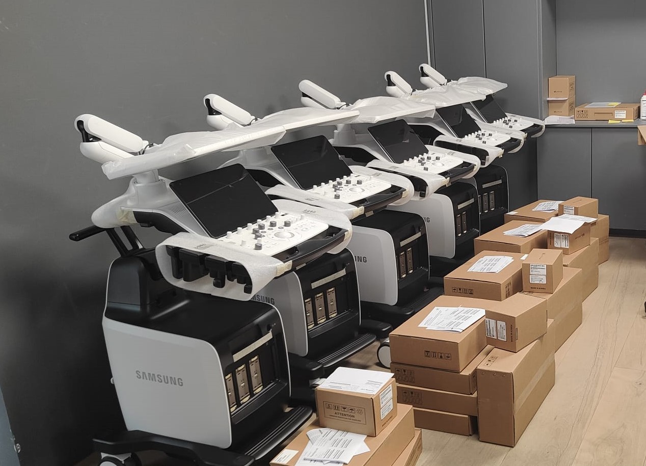
11-07-2023
We have just received the five V8s for our customer RIMED.
The devices will g...
From our calendar
Latest events from our marketing team
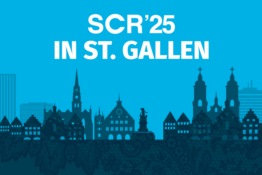
Swiss Congress of Radiology SCR'25
15-05-2025
The Swiss Society of Radiology (SGR-SSR) and its associated societies (SGPR-SSRP, SSVIR, SSER, SSSR, SSRMP), as well as the SVMTR-ASTRM (Swiss Association of Radiographers) and the SGNM-SSMN (Swiss Society of Nuclear Medicine) h
Annual Congress of the Swiss Society of Gynecology / gynécologie suisse 2025
25-06-2025

Advanced Musculoskeletal Ultrasound Course
06-09-2025
In September 2025 we are organising our Advanced Musculoskeletal Ultrasound Course in Geneva. The course combines lectures, demonstrations and hands-on trainings to teach musculoskeletal ultrasound and is desig
About us
Focusing on your Image!
We strive to bring you the best technologies for making our lives better.
- Place de l'Industrie 2, CH-1180 Rolle
- Phone: +41 22 995 01 91
- Fax: +41 22 995 01 95
- Email: meditron@meditron.ch